Table of Contents
Hallux Valgus
Hallux valgus is used to describe lateral deviation of the great toe. Metatarsus primus varus, or medial deviation of the first metatarsal with an increased first-second metatarsal angle, is commonly associated.
This deformity commonly occurs in those who wear shoes. Constrictive footwear is therefore thought to be a predisposing factor. Heredity also plays an important role in the development of Hallux Valgus (14, 15). Other associated intrinsic factors may include pes planus, Achilles tendon contracture, generalized joint laxity, hypermobilty of the first metatarso-cuneiform joint, and neuromuscular disorders.
The first MTP joint is a unique joint in the foot that has a sesamoid mechanism and a set of intrinsic muscles, which stabilize the joint. The tendons of the medial and lateral heads of the flexor hallucis brevis insert into the medial (tibial) and lateral (fibular) sesamoids, which then attach to the proximal phalanx by the plantar plate.
Three measurements are used to evaluate the presence and severity of hallux valgus:
- First metatarsophalangeal angle
- 1st-2nd intermetatarsal angle
- Sesamoid position
All are assessed on AP (dorsoplantar) view.
Radiographic Measurements
First metatarsophalangeal angle
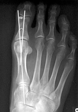
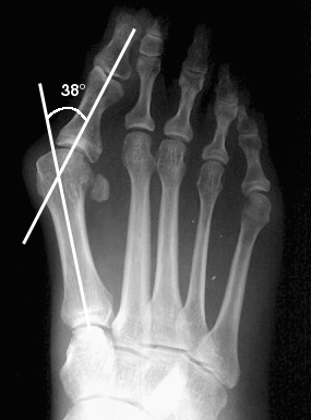
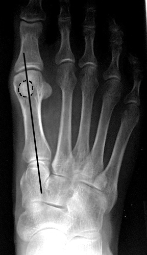
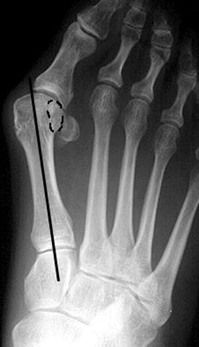
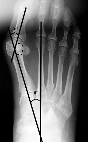
The angle is formed by a line drawn through the longitudinal axis of the first metatarsal with that drawn through the longitudinal axis of the first proximal phalanx. A normal angle is less than 15° (Fig a). Anything greater indicates hallux valgus (Fig b).
1st-2nd intermetatarsal angle
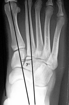
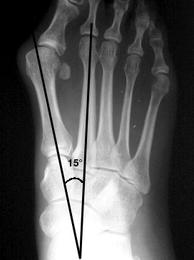
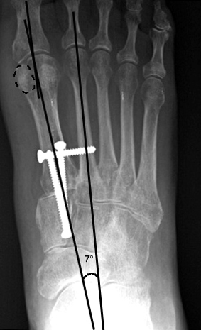
This angle is an indication of metatarsus primus varus. An angle is drawn between the longitudinal axis of the first and second metatarsal. A normal value is less than 9°. There is a close association between the degree of metatarsus primus varus and hallux valgus. The combined deformities are present to some degree in most patients (14, 15). Some believe that metatarsus primus varus is secondary to hallux valgus. However, many now believe that hallux valgus is a result of metatarsus primus varus, and surgical correction is often based on this theory.
Sesamoid position
The medial and lateral sesamoids are attached to the flexor hallucis brevis, which in turn is attached to the base of the first proximal phalanx. They are tethered to the 2nd metatarsal by the intermetatarsal ligament and by the adductor hallucis. Because of this, the sesamoids maintain a constant relationship with the 2nd metatarsal. With hallux valgus, the first metatarsal deviates medially off of the sesamoids, causing apparent lateral subluxation.
The position of the medial (tibial) sesamoid in relation to a line drawn through the mid-longitudinal axis of the first metatarsal determines sesamoid position. Traditionally there have been seven stations. Recently, a more simplified version using four stations has been developed: (16, 17)
| Station | Criterion |
|---|---|
| 0 | sesamoid completely medial to mid-axial line |
| 1 | sesamoid less than 50% overlapping the line |
| 2 | sesamoid greater than 50% overlapping the line |
| 3 | sesamoid completely lateral to the line |
Stations 0 and 1 are considered to be within normal limits (12).
The position of the tibial (medial) sesamoid of the 1st MTP joint has been used as an indication of the degree of relative subluxation of the sesamoid. However, this may be misleading. With the pull of the adductor hallucis, there is also a rotational force that pronates the great toe and 1st metatarsal, resulting in apparent subluxation on x-ray. Therefore, the degree of lateral subluxation may be overestimated.
Recent studies have shown that a tangential view of the sesamoids (“sesamoid” view) provides a more accurate assessment of of sesamoid position and degree of subluxation (17). Up to now, this has not been part of a routine foot series.
Reconstructive Surgery
A multitude of procedures for the correction of hallux valgus have been advocated. The surgical procedure most utilized by our orthopedic colleagues at Harborview Medical Center is the modified Lapidus procedure, which involves an arthrodesis of the 1st tarsometatarsal joint (18). This is often performed with a small lateral based wedge osteotomy of the medial cuneiform. This combination stabilizes the first ray and corrects the metatarsus primus varsus. Our surgeons feel that in many cases hallux valgus is a result of of a hypermobile first ray.

