| UW MSK Resident Projects |
|
|
|
|
AdamantinomaPrint-friendly version of this pagePosted by rpatel@u.washington.edu, 9/16/03 at 8:58:09 PM.
Adamantinoma of Long Bone (extragnathic adamantinoma):
Adamantinoma is a rare primary low-grade malignant tumor of bone that is locally aggressive. Adamantinoma of the long bone is not related to adamantinoma or ameloblastoma of the mandible and maxilla, which is derived from Rathke's pouch.
Demographics:
Clinical Presentation:
Approximately 60% of patients with adamantinoma have a history of trauma, including fractures, to the affected bone, but it is unclear whether trauma is involved with formation of the tumor or the formation of the tumor leaves the bone weak and susceptible to injury.
Common symptoms (duration from a few weeks to years):
· Localized, insidious aching pain · Gradual swelling and deformity of the affected limb · Limping · Increased pain with activity or lifting · Advanced or recurrent lesions may be associated with a soft-tissue mass (15%)
Imaging Findings:
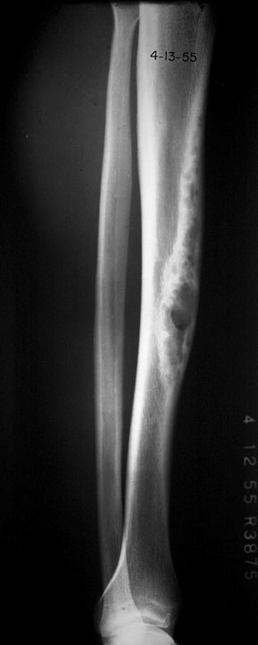
Conventional Radiographs: (Here is an example from BrighamRad)
Tibial Adamantinoma
Computed Tomography:
· Low attenuation expansile mass with sclerotic margins · Superficial erosion and soft tissue involvement
Magnetic Resonance Imaging:
MRI findings in adamantinoma are non-specific, but can help demonstrate intraosseous and extrasseous involvement.
Bone Scan (Tc-99m MDP):
Pathology:
Gross pathology:
Microscopic histology:
There are two distinctly different components:
Differential Diagnosis:
The radiographic features of adamantinoma and osteofibrous dysplasia of the tibia are generally identical.
Adamantinoma, fibrous dysplasia and osteofibrous dysplasia (ossifying fibroma) share many radiographic and pathological features, but their interrelationships are unclear. Sarisozen et al. state that there is an entity known as osteofibrous dyplasia-like adamantinoma (differentiated adamantinoma) that may be a precursor to classic adamantinoma.
Natural History:
Adamantinoma usually presents as an intraosseous, intracortical stage I-A sarcoma.
This low-grade, slowly-growing malignancy metastasizes in about 20 percent of cases by both hematogenous and lymphatic routes to other parts of the body, usually to the lungs or nearby lymph nodes. Of these cases, about 15 percent of the patients die.
Young females seem to be at the highest risk for early demise with the average age of death being 33 years compared to 48 years for men.
Treatment:
Treatment of adamantinoma is wide surgical excision - neither radiation therapy or chemotherapy is effective. Local recurrence is common if the tumor is not completely removed. Filippou et al. report a case of recurrent adamantinoma with metastasis to the lungs that was successfully retreated with surgical resection of the recurrent tumor and metastases. |
|