| UW MSK Resident Projects |
|
|
|
|
Giant Cell TumorPrint-friendly version of this pagePosted by eddiejsw@u.washington.edu, 4/26/04 at 5:40:08 AM.
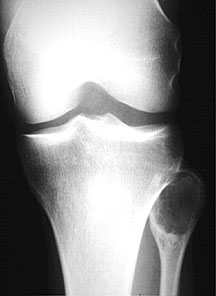
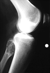
Giant Cell Tumor Introduction/Background Giant cell tumors of the bone are uncommon bone tumors which represent around 4-5% of primary bone tumors and 18% of all benign bone lesions. They are interesting in the fact that their classic radiographic appearance is easily identifiable. Giant cell tumors can either be benign or malignant, although the majority of them are benign. Radiographically, benign versus malignant tumors are difficult to distinguish. In most patients, the tumor has an indolent course, but repeated local recurrence of the tumor does happen. Approximately 5% of giant cell tumors are defined as malignant. Malignancy usually occurs as the result of malignant transformation after radiation therapy. These tumors are slightly more common in females, with 50-57% of cases occurring in females. The general age range for giant cell tumors is 20-40 years old. Approximately 85% of tumors occur in the long bones, namely the distal femur, proximal tibia, proximal humerus, and distal radius. Another location typical of giant cell tumor is the spine, particularly the sacrum. Spinal involvement is typically in the vertebral body, although its location is variable. Clinical presentation is nonspecific, and it usually is pain at the tumor site. Pathologic fracture occurs around 10% of the time. Local mass effect can cause a range of symptoms, for example, from posterior expansion into the spinal canal. Radiology: Plain film: Plain films are really the definitive imaging tool for examining giant cell tumors. The tumors are typically eccentrically located within the bone. They occur in skeletally mature patients, that is, patients with closed epiphyses. The appearance is that of a typical benign, expansile, lytic bone lesion. The location is a great discriminator, with the tumor being eccentric and adjacent to the articular surface. Although giant cell tumors are readily known as being epiphyseal lesions, early tumors can actually be found in the metaphysis only. Giant cell tumors do not have a well-defined sclerotic margin, and if there is one present, it typically is incomplete. No periosteal reaction is seen. Fig. 1 - Typical Giant Cell tumor
Spinal involvement is less specific, and vertebral body destruction and collapse can be identified. As described above, the most common location for giant cell tumor is in the vertebral body. Other imaging: CT: Imaging findings are similar to that of the plain film. MRI: Heterogeneous low-to-intermediate T1 and T2 signal for the solid portions of the tumor. Hemosiderin can be seen in 60% of the tumors. Cystic areas are of the typical high T2 signal, and fluid levels can be identified within the tumor. As is typical for MRI, it is helpful in determining the extent and local involvement of the tumor. Bone scan: Homogeneous uniform uptake, nonspecific. Treatment Typical treatment for giant cell tumors is curettage and packing of the lesions. The main complication is local recurrence. References: Dahlin, David C. Bone Tumors. Second edition. (1967). p. 78-88. Danhert, Wolfgang. Radiology Review Manual. Third edition. (1996). p.64. Resnick, et. al. Diagnosis of Bone and Joint Disorders. Second edition. (1988). Volume 6. p. 3787-3806. Weissleder, et. al. The Primer of Diagnostic Imaging. Third edition. (2003). p. 432. |
|