| UW MSK Resident Projects |
|
|
|
|
MelorheostosisPrint-friendly version of this pagePosted by braker19@comcast.net, 8/28/03 at 9:36:39 AM.
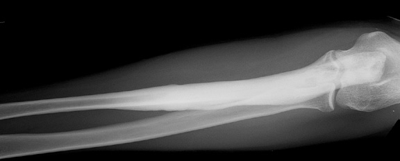
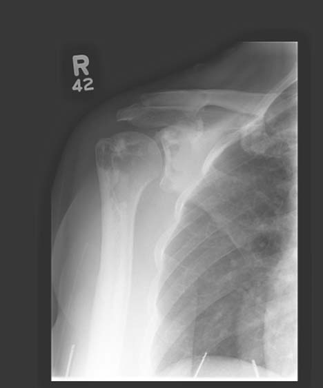
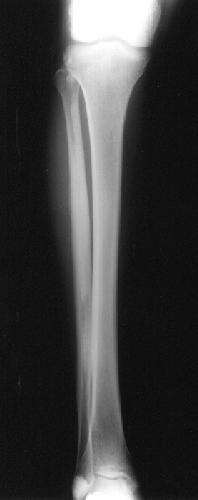
"...a rare bone disorder (of unknown etiology) whose initial manifestations include swelling of joints, pain, and limitation of motion. Eventually profound muscle contractures, tendon and ligament shortening, and soft tissue involvement with severe growth disturbances may ensue. Scoliosis, joint contracture and foot deformities may be seen." Encyclopaedia of Medical Imaging Volume III:1 Melorheostosis usually presents in a sclerotomal distribution. Early clinical findings include joint swelling, limitation of motion and pain. Contractures, ligamentous and tendinous shortening and even soft tissue involvement may ensue.(1) Radiographic findings are often limited to a single limb where more than one bone is often involved. The lower extremity is more often affected than the upper extremity. The axial skeleton is often spared. The classic finding is that of "peripherally located (cortical) hyperostosis...in one bone or a series of bones. The appearance of the osseous excrescences extending along the length of the bone simulates that of candle wax flowing down the side of the lit candle" Resnick: Bone and Joint Imaging, Second Edition. W.B Saunders Company 1996. The appearance of the lesion(s) gave the anomaly its name, which is "taken from the Greek words for member (melos) and flow (rhein). There is usually a distinct demarcation between the affected and normal bone. Dense, sclerotic linear areas are seen mainly in the cortex but also extending into the cancellous bone. It may co-exist with osteopoikilosis and osteopathia striata as well as with tumours or malformations of blood vessels or lymphatics. Soft-tissue ossifications at the site of the joint are common." (2) Other Modalities... Scintigraphy is positive and shows moderately increased uptake. Characterization can be accomplished with advanced cross-sectional imaging with Computed Tomography and MRI, but these rarely add value. The forme fruste of melorheostosis can mimic other pathology such as osteoma, myositis ossificans and parosteal osteosarcoma. Treatment includes surgical resection of soft tissue associated lesions and in extreme cases amputation may be necessary.
|
|