UW MSK Resident Projects
University of Washington, UWMC Roosevelt Clinic, Musculoskeletal Radiology

Home

About


Neurofibromatosis Type 1

Print-friendly version of this page

Posted by mchang14@hotmail.com, 1/5/04 at 3:03:06 PM.

Etiology:  Also known as von Recklinghausen disease, neurofibromatosis type 1 (NF1) is the result of a defect on Chromosome 17.

Prevalence:  1 in 3000-4000 people.  Half are caused by new mutations (mutation rate is 1 case per 10,000 population).

Inheritance Pattern:  Autosomal dominant.  100% penetrance.

Mortality/Morbidity:  Mean age of death is 54.4 years, compared to 70.1 for the general US population.  Malignant transformation of a peripheral nerve sheath tumor is 3-15%.

Diagnostic criteria (need at least 2):

  1. > 6 cafe au lait spots (larger than 5 mm in children, larger than 15 mm in adults)
  2. > 2 pigmented iris hamartomas (Lisch nodules)
  3. Axillary, inguinal freckling (Crowe sign)
  4. > neurofibroma (or 1 plexiform neurofibroma)
  5. optic nerve glioma
  6. first-degree relative with NF1
  7. dysplasia of greater wing of sphenoid

Radiologic Findings:

  1. Musculoskeletal:  Sphenoid dysplasia (empty orbit sign), pseudarthrosis, ribbon rib deformity, bowing of long bones 
  2. Spine:  Kyphoscoliosis in thoracic spine, vertebral scalloping, enlarged neural foramina, lateral thoracic meningocele
  3. Nerve sheath tumors:  neurofibromas (dumbbell lesion), plexiform neurofibroma, malignant peripheral nerve sheath tumor
  4. GI tract:  neurofibromas in stomach and bowel which may lead to obstruction/intussusception, small bowel leiomyoma, small bowel malabsorption
  5. Eye:  Lisch nodules, optic glioma
  6. Skin: cafe-au-lait spots, cutaneous and subcutaneous neurofibromas, freckling

NF1e: NF1a:  

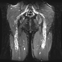
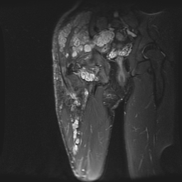
Fig 1a and b.  Two images from two different patients demonstrates extensive neurofibromas involving bilateral sciatic nerves (image a), and exiting out the sciatic notch into the peripheral nerves of the right gluteal and thigh muscles (image b).

NF1c: NF1b:  

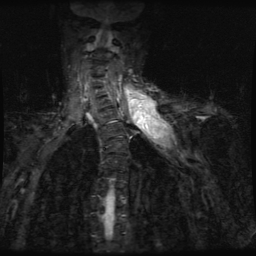
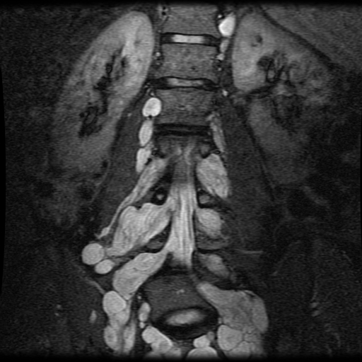
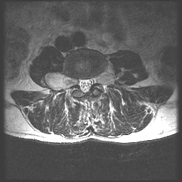
Fig 2.  Two images from the same patient shows a large paravertebral soft tissue mass engulfing C6-8 nerve roots.  Axial image demonstrates tumor extending out the left neural foramen.

NF1f: NF1g:  

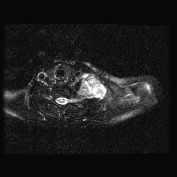
Fig 3.  Two images from the same patient demonstrates large bilateral neurofibromas involving bilateral lumbar nerve roots, and enlarging the neural foramen.  Axial image demonstrates the so-called dumbbell lesion. 

Discuss

This page was last updated: Thursday, August 4, 2005 at 12:03:05 PM
Copyright 2005 UW MSK Resident Projects
Create your own Manila site in minutes. Everyone's doing it!

This site is using the Default theme.