UW MSK Resident Projects
University of Washington, UWMC Roosevelt Clinic, Musculoskeletal Radiology

Home

About


Pigmented Villonodular Synovitis

Print-friendly version of this page

Posted by slogerfo@u.washington.edu, 5/3/04 at 8:46:29 AM.

PIGMENTED VILLONODULAR SYNOVITIS

WHAT IS IT?

  • Uncommon disease characterized by hyperplastic synovium, large effusions and bone erosions.
  • Benign lesions involving synovium membrane of joints and tendon sheaths.  Giant cell tumor of the tendon sheath of the hand and foot is considered a form of PVNS.
  • Characterized by fibrous stroma, hemosiderin deposition, histiocyte infiltrate and giant cells.
  • Etiology remains unknown but leading theory is that it is an inflammatory reaction of the synovium.
  • Two types of PVNS can occur: localized nodular form (predominantly hands and feet) or a diffuse form of the synovium.

CLINICAL PRESENTATION:

  • Incidence is approximately 1.8 cases/1 million people per year.
  • No environmental, genetic or occupation predilection.
  • Generally occurs in patients between 20 and 45 years of age.
  • Insidious onset and non-specific presentations make it a diagnostic challenge.
  • Typically monoarticular increased swelling and pain, although there are rare cases of polyarticular involvement.    
  • Joint effusion, occasionally a palpable diffuse synovial mass,  and joint stiffness.
  • Approximately 50% report an antecedent history of trauma.
  • Knee is the most commonly affected joint , followed by the hip, ankle, and small joints of hands and feet, shoulder and elbow.     
  • Aspiration of the joint yields blood-stained fluid from hemorrhage.              

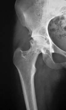
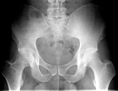
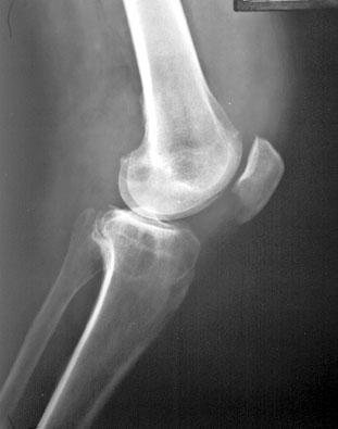
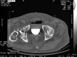
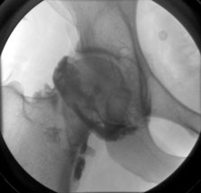
IMAGING FINDINGS:

Radiographs may be entirely normal or show minimal and non-specific findings. 

Classic radiographic appearances include bone erosions and single or multiple juxta-articular subchondral "cysts" and are commonly seen on both sides of the joint. 

Right Hip and acetabulum:

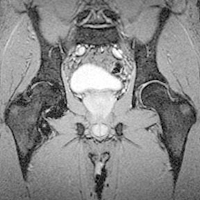
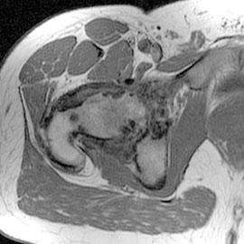
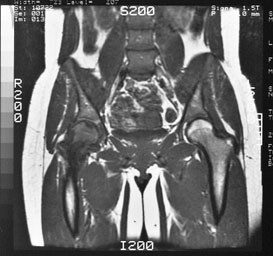
In the hip, bone lesions typically develop at the junction of the femoral head and neck, and acetabulum. Additionally, erosions are more commonly seen than in the knee due to the tight capsule of the hip joint.

Bilateral Hips:

PVNS Knee:

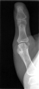
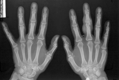
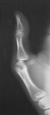
In the bones of the hands and feet, where the localized nodular form prevails, the cysts appear as sharply marginated radiolucent defects in multiple bones. 

Localized nodular PVNS in finger:

PVNS Hands:

PVNS Finger:

Giant Cell Tumor of the tendon sheath in the wrist and toe is considered PVNS of the tendon sheath

GCT wrist:

GCT toe:

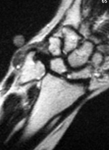
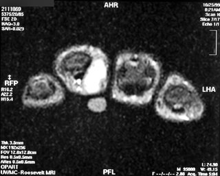
MRI is highly sensitive and specific for PVNS.  Characteristic findings include joint effusion, low signal on both T1 and T2, and gradient echo secondary to hemosiderin deposition, hyperplastic synovium, bone erosions and preservation of bone density.

Hips coronal MRI GRE:

Hip axial T1:

Hips coronal T1:

On CT, the cystic erosions appear as decreased signal of the marrow.

Hips axial CT:

Arthrography is another imaging modality that can be used in the diagnosis of PVNS and may show a richly vascular process,increased articular cavity, joint effusion or synovial villae.

Arthrogram hip:

Bone scan with Tc Pertechnetate may be useful for detecting inflammation of the synovial membrane.

DIFFERENTIAL DIAGNOSES:

  • Osteoarthritis
  • Rheumatoid Arthritis
  • Gouty Arthritis
  • Psoriatic Arthritis
  • Amyloidosis
  • Tuberculous Arthritis
  • Synovial chondromatosis
  • Synovioma
  • Systemic lupus erythematosus
  • Avascular Necrosis (hip only)

TREATMENT:

  • Localized form is best treated by local excision of the nodular lesion.
  • Synovectomy with curretage of osseous lesions is performed for diffuse form.
  • Due to relatively high rate of local recurence, some have advocated adjuvant radiation therapy.

REFERENCES:


slogerfo@u.washington.edu


Discuss

This page was last updated: Thursday, August 4, 2005 at 11:23:17 AM
Copyright 2005 UW MSK Resident Projects
Create your own Manila site in minutes. Everyone's doing it!

This site is using the Default theme.