| UW MSK Resident Projects |
|
|
|
|
Radiation Changes to BonePrint-friendly version of this pagePosted by moraske@yahoo.com, 2/3/04 at 7:42:23 PM.
Overview There are two main reasons that bones receive radiotherapy: 1) an intentional delivery to bones to treat painful metastatic bone lesions or 2) their unavoidable inclusion into a radiation field which intends to treat an adjacent soft tissue neoplasm. In either case, the effects on the bone are varied and depend upon: 1. Dosage. 2. Quality of the x-ray beam. 3. Age of the patient. 4. Method of fractionation (fractionating treatment enables the physician to give the patient the necessary treatment dose while separating its delivery by a time interval in order to reduce toxic side effects to normal tissue). 5. Length of time of therapy. 6. Specific bone or bones involved. 7. Existence of trauma or infection at the site.
The main effects on the bone include: disruption of normal growth and maturation, scoliosis, osteonecrosis, and neoplasm formation. Growth arrest and disturbances Growth arrest, in general, is believed to be caused by a combination of cellular injury to chondrocytes and damage to the physeal blood vessels. The epiphysis is the most sensitive area to radiation and it has been shown that delivery of a total of 20 Gy to immature bone can cause growth disturbance. Radiotherapy can also increase the risk for epiphyseal plate trauma such as slipped capital femoral epiphysis. Radiation delivered to the metaphysis can cause bowing, fraying and sclerosis and look similar to rickets. The diaphysis is thought to be relatively more radioresistant and periosteal bone formation may be less affected than enchondral bone formation. Nevertheless, when an entire bone is exposed to radiation, it will demonstrate narrowing of the shaft and an overall reduction in size and diameter.
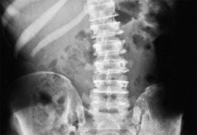
Hypoplastic pelvis and lumbar scoliosis in a patient who received radiotherapy as a child.
Scoliosis As with bone growth abnormalities in other areas of the body, the reason for radiation induced scoliosis are multifold. In general, scoliosis induced by radiotherapy is thought to be mild and non progressive. It affects the growing spine more severely than mature spine. Children who are treated before the age of 2 show the largest degree of abnormality. Uneven growth arrest, leading to curvature, was initially thought to be due to "fall off" at the edge of the x-ray beam and some investigators believed that irradiating the entire vertebral body equally would prevent scoliosis from developing. This has not shown to be true, although asymmetric radiation is associated with more severe and more frequent deformities. Changes can occur with doses of 10 Gy and higher and generally occur 9 to 12 months after treatment.
Osteonecrosis In a mature skeleton, "growth damage" manifests as osteonecrosis. Radiation necrosis is dose-dependant, with "radiation osteitis" seen at around 30 Gy and osteoradionecrosis assoicated with doses of 50 Gy or higher. Radiation osteitis is a term used to describe potentially reversible changes such as temporary cessation of growth, periostitis, bone sclerosis and increased fragility, ischemic necrosis and infection. Radiographs will show bone which is mottled demonstrating both osteopenia and sclerosis and areas of coarse trabeculation. Some investigators and clinicians believe that radiation osteitis is the set-up for osteoradionecrosis and that a patient does not progress to necrosis unless infection is present. Others favor a "cell death" theory. Recent evidence suggests that radiation induces osteoblasts to enter G2 cell cycle arrest and sensitizes them to agents which mediate their apoptosis. Radiation induced vascular injury is also a likely culprit to the development of radiation osteitis and osteonecrosis. Its role, however, is debated. Changes are seen at different time intervals depending on which bone is irradiated. There are several bones which have characteristic complications which are listed below. Mandible: Frequently the changes are seen within a year of treatment. It is a more superficial bone, and therefore it inadvertently receives a larger dose during treatment of head and neck neoplasms. One of the major concerns with osteoradionecrosis of the mandible is that can be very difficult to differentiate it from tumor recurrence. Pain, ulceration, and bleeding can be present, but do little to determine the diagnosis as these symptoms are common to both entities. Pelvis: Avascular necrosis of the femoral head can, of course, be seen. In 2% of patients, fractures of the femoral head occur. Protrusio acetabuli is also a reported complication. Femoral neck fractures have been seen as early as 5 months after therapy and with as little as 16 Gy of radiation given. The fractures generally heal with normal callous formation. The SI joints may also be effected showing bilateral, symmetric sclerosis. Shoulder: Frequently in the treatment of breast carcinoma, the shoulder receives radiation. This can induce osteopenia and/or a coarse and disorganized pattern of trabeculation which looks like Paget's disease. Rib, clavicular and scapular fractures are seen. In some cases, there can be avascular necrosis of the humeral head. Sternum: Pectus excavatum can develop. Other complications include osteopenia, disorganized trabecular pattern, and necrosis involving one or more sternal segments.
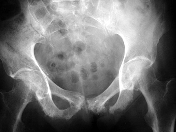
Osteonecrosis of the pubic rami Radiation-induced neoplasms Radiation-induced tumors in patients who receive radiotherapy before 2 years of age are usually benign. The most commonly reported tumor in this group of patients is osteochondroma, and are thought to be induced with doses ranging from 16-64 Gy. In patients who received radiotherapy during adulthood, induced tumors are more likely to be malignant. Osteosarcoma and fibrosarcoma are generally considered to be the most common radiation-induced tumors in this population. Chondrosarcoma and malignant fibrous histiocytoma are also seen. Specific criteria aid in diagnosing a radiation-induced sarcoma and are as follows: 1. The sarcoma arises in an irradiated field. 2. There is a 4 year latent period 3. The sarcoma is histologically different from the original tumor and radiation was delivered in the absence of a diagnosis of the sarcoma. Radiation-induced sarcomas have been shown to develop anywhere from 4 to 42 years after therapy. The average latent period is 11 years. The radiation doses associated with tumor development vary, but usually at least 30 Gy is needed. As mentioned previuosly, tumor can sometimes be confused with osteonecrosis, as they can demonstrate similar symptoms. In those cases, serial radiologic exams or biopsy may be needed.
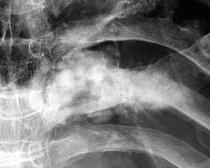
Osteosarcoma of the rib in a patient who had received radiation therapy for lung carcinoma 9 years earlier
|
|


