| UW MSK Resident Projects |
|
|
|
|
Scapholunate InstabilityPrint-friendly version of this pagePosted by wangdo@u.washington.edu, 8/15/03 at 2:51:48 PM.
Carpal Instability
Definition
The wrist, with its enormous range of motion, is one of the most complex and frequently used joints within the human body. However, because of the prominent role it plays in normal daily activities, the wrist can oftentimes wind up being a source of significant disability and pain.
The proximal bones of the wrist (scaphoid, lunate, and triquetram) have no muscular attachment. As a result, their main purpose is to stabilize the wrist joint and provide a focal plane to help transmit the force generated by the flexors and extensors of the arm.
To help them in this task, numerous strong ligaments bind the proximal bones together, allowing them to function more cohesively. Damage to any of these ligaments results in either acute symptoms or a less noticeable but possibly more dangerous malalignment of the carpal bones that, if left uncorrected, will accumulate abnormal wear and tear -- eventually leading to severe and debilitating osteoarthritis.
Ligamentous injuries and subsequent carpal instability tend to occur in young and middle aged patients. This is because as people age, their bones become more fragile and thus trauma in the elderly tends to cause outright bone fractures instead of soft tissue damage. The most frequently encountered ligamentous injury of the wrist is scapholunate instability.
Three main ligaments bind the Lunate and Scaphoid bones together:
· Volar radioscapholunate ligament · Scapholunate interosseous ligament · Dorsal scapholunate ligament
Scapholunate instability is defined as significant injury to two or more of these ligaments.
Cause
Scapholunate instability is usually caused by stress placed on a hyperextended wrist, especially in ulnar deviation (such as frequently happens when patients fall on an outstretched hand). When this occurs, the ligaments binding the scaphoid and lunate bones begin to tear, starting with the scapholunate interosseous ligament.
When a ligamentous wrist injury occurs, patients will generally describe one or more of the following symptoms:
One of the easiest ways to evaluate these patients is with a simple physical exam maneuver known as the Watson test.
Watson Test
Watson HK, Ashmead D4, Makhlouf MV: Examination of the scaphoid. J Hand Surg Am. 1988; 13: 657-660.] A painful snap is obtained when pressing hard on the scaphoid tubercle volarly and moving the wrist from ulnar to radial deviation, the wrist being maintained in axial compression. If clinical suspicion is high, further evaluation with imaging studies can be done.
Radiologic Diagnosis
As the ligaments binding the scaphoid and lunate bones are damaged, the scaphoid begins to move out of its normal position and rotationally sublux. This leads to several noticeable findings on radiographic examination.
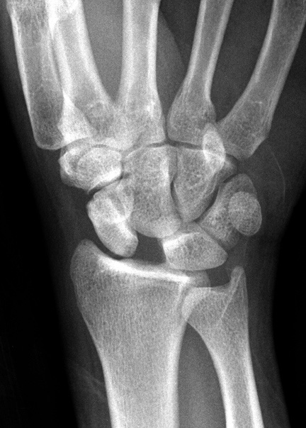
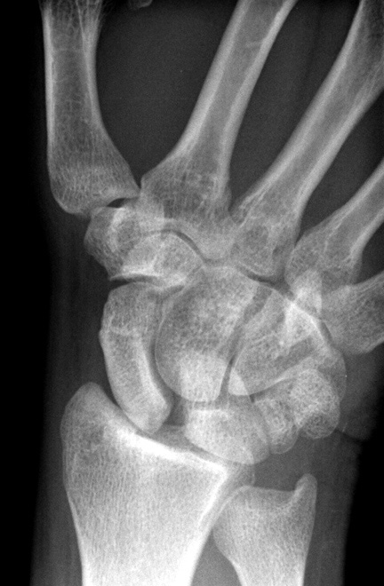
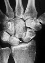
On AP plain films of the wrist, an abnormally large gap between the scaphoid and lunate bones can oftentimes be seen. This is known as the Terry Thomas sign, in reference to the British comedian with a sizeable gap between his two front teeth (or David Letterman or any other person with a gap in their teeth, sign).
AP Wrist
Generally, a scapholunate interval of > 2mm is suspicious and > 4 mm is virtually diagnostic for scapholunate dislocation or instability. Lateral WristLateral X-rays of scapholunate instability may also demonstrate an increased capitolunate angle > 15 degrees as well as increased scapholunate angle > 60 degrees.
Arthrography While fairly sensitive, plain films will sometimes fail to see the early signs of scapholunate instability when the ligaments are torn but the scaphoid has yet to sublux. In these situations when suspicion is high, or when plain films have already detected the dislocation and further evaluation is needed to plan treatment, arthography is the next logical step in radiologic evaluation. The wrist can be divided into three discrete compartments with ligamentous borders that prohibit the movement of fluid from one compartment to the next. The scapholunate ligaments comprise part of this border and any tear can be diagnosed on arthrography when contrast injected into one compartment can be seen abnormally flowing into a neighboring compartment.
However, while often diagnostic, contrast leakage does not always signal a significant tear within the ligaments but instead may only represent a ligamentous perforation without significant consequences. If this is suspected to be the case, contrast injection can be coupled with CT scan or MRI of the joint to directly assess the health of the carpal ligaments. Furthermore, CT scan or MRI can directly determine the site and length of ligament tears and is a critical tool when planning any surgical repair.
Treatment
Because scapholunate injuries can sometimes present without symptoms yet if left untreated can eventually result in further carpal derangement and debilitating osteoarthritis, prompt diagnosis and repair is extremely important.
If the injury is thought to be minimal, conservative treatment with splinting and NSAIDS can be used to provide symptomatic relief. However, any significant degree of injury necessitates surgical correction.
The overall aim of surgery is to restore a normal orientation of the scaphoid with the proximal pole appropriately articulating with the radius and to correct any other abnormalities that have occurred if the original injury was undiagnosed and uncorrected.
Numerous surgical techniques have been described, but generally revolve around soft tissue repair, where the carpal ligaments are modified or restored, or limited carpal arthrodesis, where the wrist bones are either orthopedically fixed or fused.
If scapholunate insufficiency is left untreated long enough, further shifts of the carpal bones can occur, eventually leading to a syndrome known as scapholunate advanced collapse, or SLAC:
As the scaphoid moves further from the lunate, the normal load-bearing ability of the capitolunate joint disappears. Among other things, this results in the migration of the capitate bone proximally, driving a further wedge between the scaphoid and lunate.
Check out the following scapholunate instability case in John Hunter's file. Another example of scapholunate ligament tear seen on arthrography can be seen in this case. |
|