| UW MSK Resident Projects |
|
|
|
|
Posterior Sternoclavicular DislocationPrint-friendly version of this pagePosted by passm@u.washington.edu, 2/27/04 at 3:31:48 PM.
POSTERIOR STERNOCLAVICULAR DISLOCATION What is it? An unusual dislocation at the sternoclavicular joint that is usually the result of compressive forces on the shoulder girdle. The forces of posterior compression transmit throught the fulcrum of the costoclavicular ligament and result in posterior dislocation. Alternatively, a direct blow to the anteromedial clavicle can result in posterior dislocation.
Who gets it? Clasically described in American football players who get "dog piled" while in a lateral position. Our case happens to be a rugby player who sustained a tackle injury. It can certainly happen in a MVC when the forces are just right.
Who cares? People who sustain this unusual injury can also sustain some pretty serious associated injuries. These include: 1. Direct trauma to the great vessel origins, or brachiocephalic vein. Mediastinal hematoma is usually present. 2. Respiratory distress from compression on the trachea. 3. Voice change from compression of the recurrent laryngeal nerve. 4. Pneumothorax
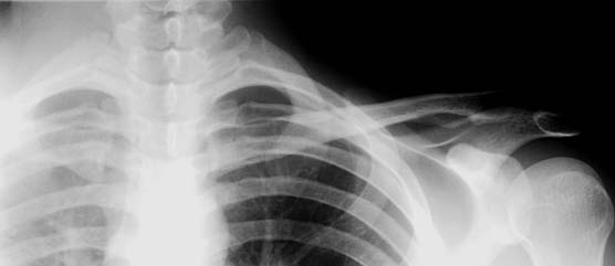
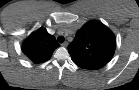
How do we see it? Special plane film views are described, but few people know these, and even fewer can read them. CT is the modality of choice because it deliniates the anatomy of the joint so well. It also demonstrates the degree of separation, and can also show additional diagnoses. (see associated injuries) If clinical suspicion is high... go directly to CT. Our Case: A young rugby player who sustained an injury (takcle?), and complained of severe sternum pain. Interestingly, he later complained of some dysphagia.
This film is nice to have, but does not help us alot. We need CT.
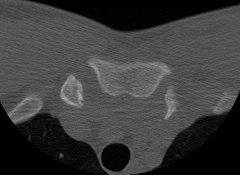
That is a scary looking bony protuberance pointing posteriorly on the left. Once you see the post contrast it is easier to see why.
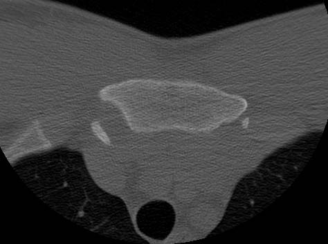
A small avulsion fragment.
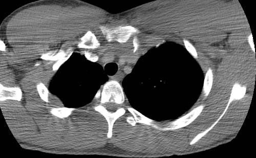
Very close to the brachiocephalic vein. The great vessels look ok.
Anterior mediastinal hematoma. Our patient underwent closed reduction of the dislocation with good results. He did not have one of the serious complications listed above. I guess the dysphagia resolved.
REFERENCES: |
|