| UW MSK Resident Projects |
|
|
|
|
Septic arthritisPrint-friendly version of this pagePosted by wbrinkma@u.washington.edu, 4/7/04 at 1:13:36 PM.
Septic Arthrits
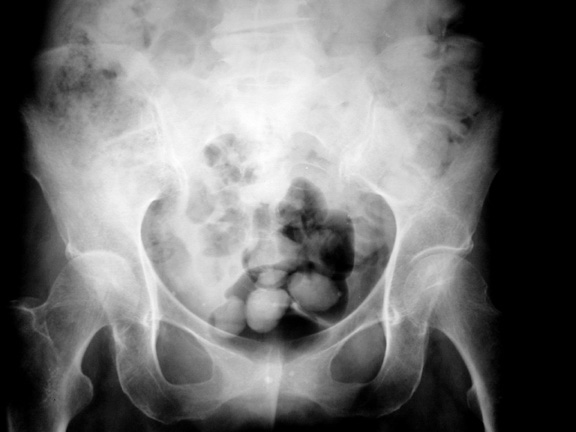
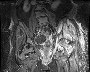
Septic arthritis is a common destructive arthropathy that requires prompt diagnosis and treatment to avoid the significant morbidity that results from the loss of a joint. Therefore, it should be strongly considered in the differential diagnosis of monoarticular disease. Traditionally, the diagnosis of septic arthritis was based on clinical assessment and prompt arthrocentesis. However, the clinical picture may be obscured by multiple confounding factors and a paucity of specific findings especially for the deep joints, ie. the hip or shoulder. Imaging can be used to confirm the diagnosis of septic arthritis and more importantly, imaging findings suggestive of septic arthritis can direct the clinician to a diagnosis that may not have been considered. Plain film findings of septic arthritis include: joint effusion, soft tissue swelling, periarticular osteoporosis, loss of joint space, marginal and central erosions and bone ankylosis. CT is more sensitive than plain films for the detection of early bone destruction and effusion. The role of MRI in the diagnosis of septic arthritis has been increasing in recent years in an effort to detect this entity earlier. Findings are usually evident within 24 hours following the onset of infection and include: synovial enhancement, perisynovial edema and joint effusion. Signal abnormalities in the bone marrow can indicate a concomittant osteomyelitis. The sensitivity and specificity of MRI for the detection of septic arthritis has been reported to be 100% and 77% respectively.
Septic arthropathy right hip with joint space loss and loss of the subchondral line in the tectum of the acetabulum
Coronal STIR-weighted sequence of the hips demonstrates a large left joint effusion with perisynovial edema. Abnormal marrow hyperintensity within the proximal femur and acetubulum consistent with concomittant osteomyelitis
References 1. Karchevsky M, Schweitzer ME, Morrison WB, Parellada JA. MRI findings of septic arthritis and associated osteomyelitis in adults. AJR 2004; 182:119-122. 2. Resnick D. Bone and joint imaging. Philadelphia, PA: WB Saunders Co; 1989; 744-749 3. Stoller DW, Tirman P, Bredella MA. Diagnostic imaging orthopaedics. Salt Lake City, UT: Amirsys; 2004; 4-99. |
|