Table of Contents
Pes Planus
Theory of Development of Pes Planus
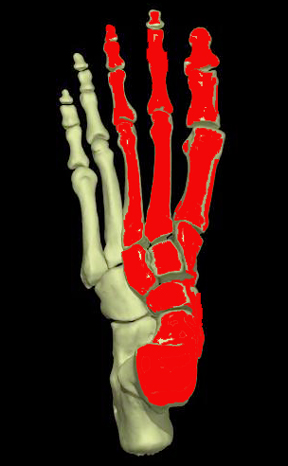
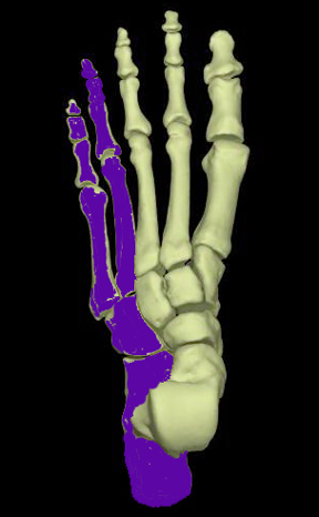
The foot can be divided into two major parts, the medial column and the lateral column. The talus, navicular, cuneiforms, and the first three rays comprise the medial column and the calcaneus, cuboid, and lateral 2 rays comprise the lateral column (5). The lateral column is inherently stable. The medial column has an adaptive function during the weight-bearing stage and acts as a stabilizer during the propulsive phase.
(from Abrahams PH. Interactive Skeleton (CD-ROM). Mosby Multimedia, 1995)
Although the etiology of symptomatic adult flatfoot is still debated, most agree that it is produced by abnormal repetetive loading on the medial column, which leads to attenuation or dysfunction of the ligamentous and tendinous stabilizers of the medial column, resulting in the bony deformities seen both clinically and radiographically. Thus, although the inciting factor leading to the development of flatfoot is not well understood, it is clear that injury to both dynamic and static structures in the foot are responsible for the deformity we call flatfoot.
The severity of flatfoot deformity on radiographs does not correlate well with symptoms, i.e.; some cases with extensive radiographic deformities may be asymptomatic whereas other cases of mild deformity may be significantly symptomatic. In addition, the onset of flatfoot deformity is often insidious. Thus, it is important to be able to recognize early radiographic signs. This variability in presentation underscores the need to take into account all available information, both clinical and radiographic, when evaluating foot alignment abnormalities.
Radiographic Measurements (List of All Measurements)
Radiographic Findings of Symptomatic Adult Flatfoot (Pes planus)
Clinically and radiographically, symptomatic adult flatfoot is a complex abnormality involving all three dimensions and multiple joints within the foot (6). Each measurement used to assess flatfoot is a two dimensional representation of this three-dimensional abnormality (7). Therefore, one must take into account all information available on both views in making an overall assessment of alignment.
There are basically 3 components that are involved in producing the alignment abnormalities of symptomatic adult flatfoot:
- collapse of the longitudinal arch
- hindfoot valgus
- forefoot abduction
Each of these components can be assessed on either the lateral or AP view of the foot.
The most useful measurements are in bold:
FOREFOOT ABDUCTION
- AP: Talonavicular coverage angle
- AP: 1st metatarsal talar angle
COLLAPSE OF LONGITUDINAL ARCH
- Lateral: 1st metatarsal talar angle
- Lateral: Calcaneal pitch
HINDFOOT VALGUS
- Lateral: Talo-calcaneal angle
- AP: Talo-calcaneal angle
OTHER SIGNS
- AP & Lateral: CYMA line
Radiographic Work-up (Most Useful Measurements)
Talonavicular coverage angle
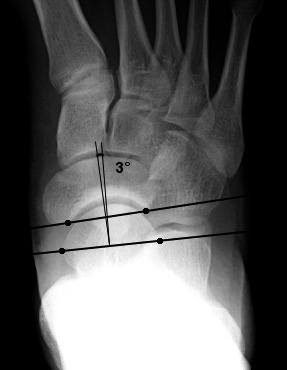
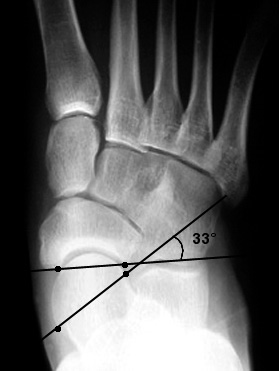
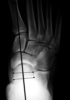
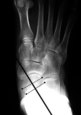
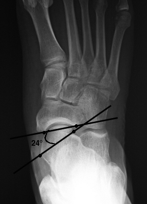
A measurement that is quite useful for evaluating pes planus on AP views is lateral subluxation of the navicular on the talus, or talonavicular uncoverage (2, 8). This is an indication of forefoot abduction, one of the three components of flatfoot. This measurement is taken off a weight-bearing AP (dorsolateral view). This angle represents the degree of shift of the navicular on the talus. Two lines are drawn, one connecting the edges of the articular surface of the talus, and one connecting the edges of the articular surface of the navicular. The angle formed by these two lines is the talonavicular coverage angle (Fig a). An angle of greater than 7 degrees indicates lateral talar subluxation. (Fig b)
Normal and Abnormal Talonavicular Angle
Lateral talar - 1st metatarsal angle
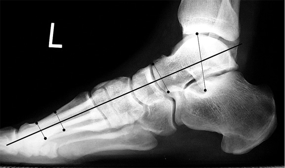
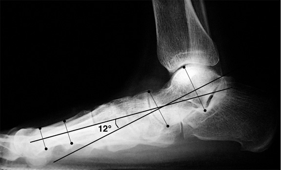
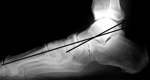
Probably the most familiar line to radiologists, and a more direct measurement of pes planus, or collapse of the longitudinal arch, is the talar-1st metatarsal angle (Meary's angle)(3). This is an angle formed between the long axis of the talus and first metatarsal on a weight-bearing lateral view (Fig a). This line is used as a measurement of collapse of the longitudinal arch. Collapse may occur at the talonavicular joint, naviculo-cuneiform, or cuneiform-metatarsal joints.
In the normal weight-bearing foot, the midline axis of the talus is in line with the midline axis of the first metatarsal. An angle that is greater than 4° convex downward is considered pes planus (Fig b) with an angle of 15° - 30° considered moderate , and greater than 30° severe (8, 9). An angle greater than 4 degrees convex upward is considered a pes cavus(10, 11).
a. Normal Meary's angle. The long axis of the talus intersects that of the first metatarsal.
b. The long axis of the talus is angled plantarward in relation to the first metatarsal, consistent with pes planus.
Calcaneal pitch
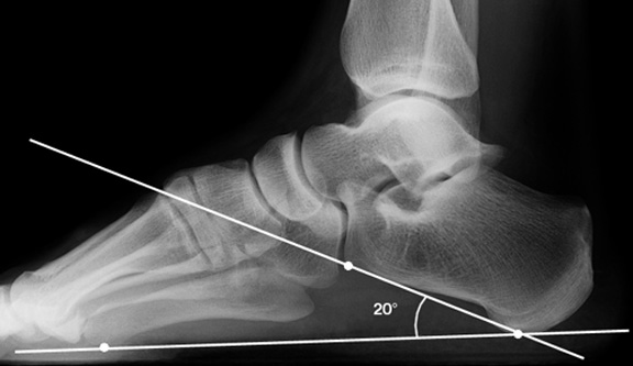
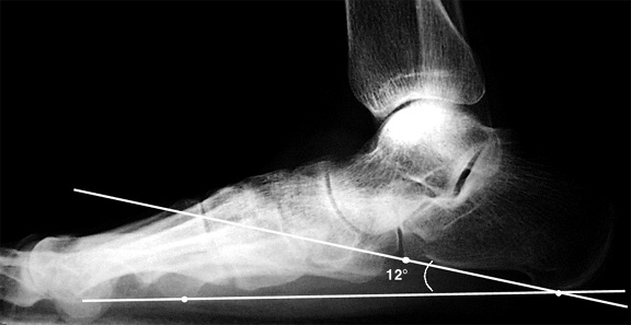
A line is drawn from the plantar-most surface of the calcaneus to the inferior border of the distal articular surface. The angle made between this line and the transverse plane (or the line from the plantar surface of the calcaneus to the inferior surface of the 5th metatarsal head) is the calcaneal pitch (Fig a)(5, 12). A decreased calcaneal pitch is consistent with pes planus.(Fig b) Unfortunately, there have been differing opinions between authors concerning the normal range of calcaneal pitch 18 to 20°is generally considered normal (12), although measurements ranging from 17 to 32° have been reported to be normal (5).
a. Normal calcaneal pitch.
b. Decreased calcaneal pitch indicating pes planus.
Other Measurements
AP Talar - 1st metatarsal angle
This is another method for evaluating the degree of midfoot and forefoot abduction. A line drawn through the mid-axis of the talus should be in line with the first metatarsal shaft, If it is angled medial to the first metatarsal it indicates pes planus.
Lateral Talocalcaneal Angle
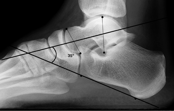
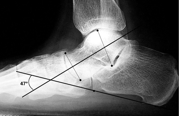
The lateral talocalcaneal angle is the angle formed by the intersection of the line bisecting the talus with the line along the axis of the calcaneus on lateral weight-bearing views. A line is drawn at the plantar border of the calcaneus (or a line can be drawn bisecting the long axis of the calcaneus).The other line is drawn through two midpoints in the talus, one at the body and one at the neck. The angle is formed by the intersection of these axes. The normal range is 25-45 degrees (Fig a). An angle over 45 degrees indicates hindfoot valgus, a component of pes planus (Fig b) (5,13).
a. Normal lateral talocalcaneal angle.
b. Increased talocalcaneal angle indicaitng hindfoot valgus in pes planus.
AP Talocalcaneal angle (Kite's angle)
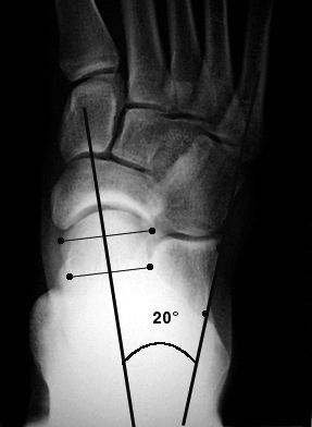
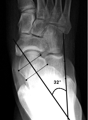
AP talocalcaneal angle (Kite's angle)tends to be unreliable and difficult to measure because many AP radiographs not well exposed in this region. This is the angle formed by the intersection of a line bisecting the head and neck of the talus and a line running parallel with the lateral surface of the calcaneus. The range of normal for adults is 15 - 30° (Fig a) (14). An angle greater than 30° would indicate hindfoot valgus, seen with pes planus (Fig b).
CYMA line
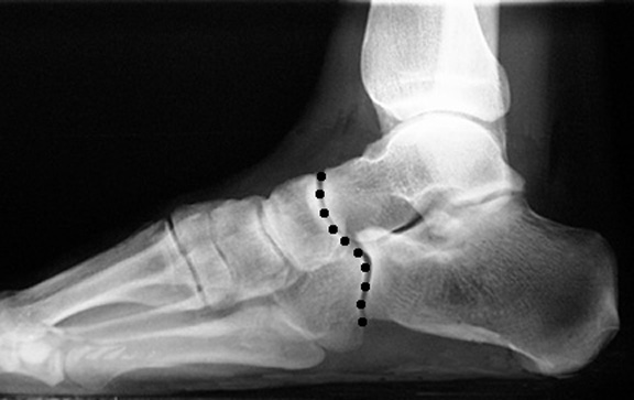
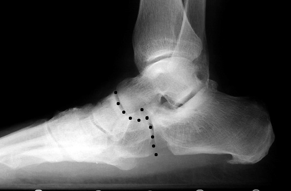
A cyma line is an architectural term designating the union of two curve lines. A normal midtarsal joint should create a smooth cyma between the talonavicular joint and calcaneocuboid joint on both the AP and lateral views (Figures a). If the cyma line is broken it suggests “shortening” of the calcaneus relative to the talus (Figures b). This is often just a radiographic shortening possibly due to rotation of the talus on calcaneus (typically seen in a patient with adult flatfoot including loss of the medial arch). It may, however, be due to actual shortening of the calcaneus and some surgeons would argue that a lateral column lengthening should be performed in addition to a medial column stabilization.
AP CYMA Line
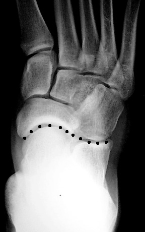 | 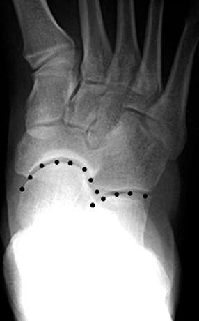 |
| a. Normal AP cyma line: Line between talonavicular joint and calcaneocuboid joint is smooth and continuous. | b. Broken cyma line. |
Lateral CYMA Line
a. Line connecting talonavicular joint and calcaneocuboid joint is smooth and continuous.
b. Broken Cyma line of pes planus.
Reconstructive Surgery for Pes Planus
There are two main options for surgical reconstruction utilized by our Orthopedic surgeons at Harborview Medical Center: the medial column stabilization and lateral column lengthening procedures.
The advantages of these two procedures is that the essential joints of the foot necessary for adaptive mobility (ankle, subtalar, and talonavicular joints) are retained.
Medial column stabilization
Medial column stabilization, used when there are predominant mid and forefoot abnormalities, is an arthrodesis of the 1st metatarsal cuneiform joint and/or the naviculocuneiform joint.
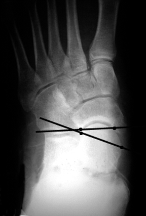
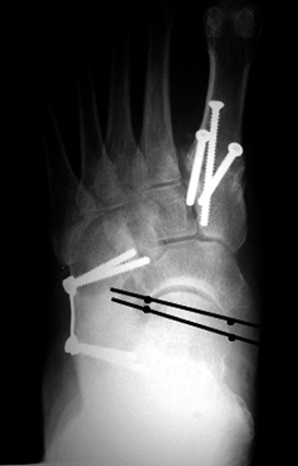
 | 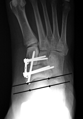 |
| a. AP view showing lateral peritalar subluxation. | b. Postoperative view after medial column stabilization showing correction of this talonavicular uncoverage. |
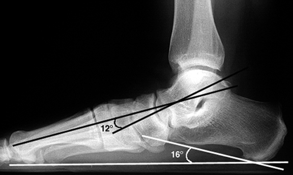
a. Abnormal lateral talar-1st metatarsal angle (black) and calcaneal pitch (white) indicating pes planus.
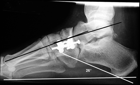
b.Post-operative view after medial column stabilization, showing correction of these angles to normal.
Lateral column lengthening
Lateral column lengthening is used is used when a medial column stabilization will not produce a powerful enough correction. It is usually performed in addition to a medial column stabilization. It involves placement of a bone graft within the calcaneocuboid joint. With both procedures, there is usually heel cord lengthening or gastrocnemius recession, and transfer of the flexor digitorum into the medial cuneiform or stump of the posterior tibialis tendon (which is usually torn).
a. Abnormal lateral talar-1st metatarsal angle (black) indicating pes planus.
b. Post-operative lateral view after lateral and medial column stabilization, showing correction of these angles to normal.

