Anatomy and Pathology
Interactive CT Anatomy Learning Module | University of Washington
| Home | Congenital | Inflammatory | Trauma | Tumor | Interactive Atlas | Quiz |
University of Washington
Seattle, Washington |
Introduction to Interactive Temporal Bone Anatomy |
|
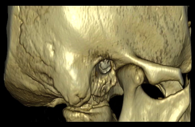
Temporal bone anatomy is complex, and further complicated by the small size and three-dimensional orientation of associated structures. Computed tomography (CT) has revolutionized imaging of the temporal bone. Recent advances in 32, 64 and now 128-slice CT scanners allow the acquisition of high-resolution, volumetric data that allows image reconstruction in any plane. In addition to the traditional axial and coronal planes, it has become routine to also obtain reconstructions in the Stenvers and Poschl projections. The Stenvers projection is the plane parallel to the long axis of the petrous bone and the Poschl projection is the plane parallel to the short axis of the petrous bone. These additional planes are extremely useful for evaluating the structures of the middle and inner ear that may not be as well seen on the standard axial and coronal planes. A wide array of pathology may affect the relatively small anatomical region of the temporal bone. Often there is overlap of clinical symptoms in pathological entities arising from the temporal bone. Thus, the combination of a good clinical history in conjunction with a dedicated temporal bone CT facilitates evaluation of the temporal bone for subtle or not-so subtle disease. This interactive web-based temporal bone anatomy module provides the user an opportunity to review normal temporal bone anatomy and abnormal cases involving four general categories of pathology affecting the temporal bone: congenital malformations, inflammatory conditions, trauma and tumor and tumor-like conditions. There is also a self-assessment quiz with questions pertaining to these four general categories. |