
TMJ CT
UW Radiology Home
TMJ Tutorial
Diagnostic Radiology Anatomy Modules
TMJ CT
Computed tomography (CT) can be used to diagnose internal derangement and other disorders of the TMJ. The patient is scanned in either the transverse or direct sagittal plane using thin sections (1-2 mm) and a soft tissue technique. If transverse sections are obtained, sagittal reconstructions are made through the condyle. The meniscus can be visualized on CT since it is slightly higher in density than the surrounding muscle and soft tissue. Since there is only a small difference in density between the meniscus and soft tissue , then either narrow window settings or the Identity Mode[trademark] (or equivalent) must be used to identify the meniscus.
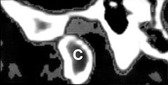
normal TMJ CT showing normal disk posterior and superior to condyle (C)
Normally, there is only a small amount of increased soft tissue density anterior to the condyle on CT. In internal derangement, the anteriorly displaced meniscus results in abnormally increased soft tissue density anterior to the condyle.
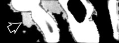
displaced meniscus (arrow) anterior to the condyle
For further information, contact Thurman Gillespy III, M.D.
© 1994 University of Washington Department of Radiology
All rights reserved. Do not use without written permission.
Last update: Thursday, May 10, 2001 at 2:27:11 PM.