of Acetabular Fractures:
Interactive CT Anatomy Learning Module | University of Washington
| Home | Quiz | Column Principle | Posterior Wall Fracture | Both Column Fracture | Transverse Fracture | Interactive Atlas | Summary | References |
| Both Column Fracture | ||||||||||||||
|
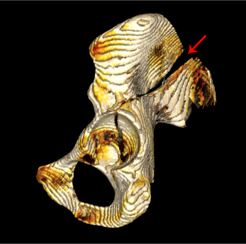
Lateral view of a 3D volume-rendered reconstruction of the hemipelvis with the femur removed. Red arrow denote a vertically-oriented fracture extending from the iliac wing through the acetabulum down into the ischiopubic ramus, resulting in free-floating anterior and posterior fragments.
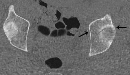 Axial CT image of the pelvis through the left acetabular tectum. Black arrows denote a coronally-oriented fracture plane through the acetabulum.
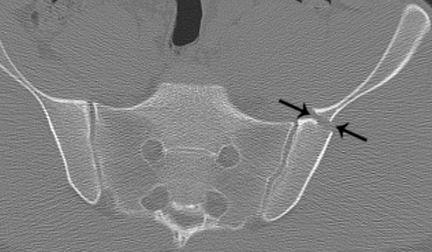 Axial CT image of the pelvis through the sacroiliac joint. Black arrows denote extension of fracture up the iliac wing.
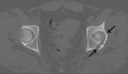 Axial CT image through the left acetabular tectum. Black arrows denote an obliquely-oriented fracture line through the posterior acetabular wall.
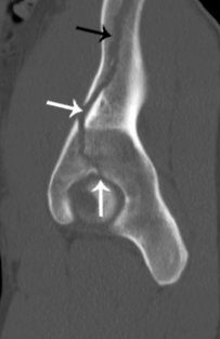 Sagittal CT image through the acetabulum, lateral to medial. White arrows denote a vertically-oriented fracture through the acetabulum and black arrows indicate extension through the anterior column up the iliac wing.
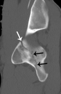 Sagittal CT image through the acetabulum, lateral to medial. White arrow denotes a vertically-oriented fracture through the acetabulum and black arrows indicate extension through the posterior column towards the ischial tuberosity. Both column fractures are the most common pattern (19 – 29%) and also the most devastating. The fracture plane travels vertically through the innominate bone, divides it anteriorly and posteriorly into at least three fragments. The fragments containing the acetabulum are free-floating, meaning they are no longer in continuity with the portion of the ilium that articulates with the sacrum. Therefore, the ipsilateral lower extremity is isolated from the axial skeleton and the fracture is unstable. On axial images through the acetabular tectum, the fracture is oriented coronally. It extends up the iliac wing and down the iliopectinal and ilioischial line into the ischiopubic ramus.
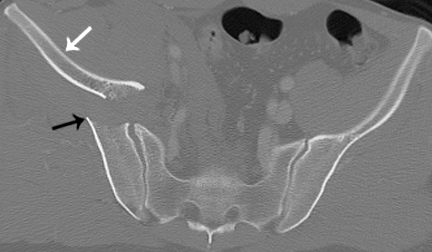
Axial image through the sacroiliac joint demonstrates the spur sign. Black arrow denotes the point of the spur and white arrow indicates the medially displaced iliac wing fragment.
When the detached iliac fragments are medially displaced, the lateral-most edge of the iliac bone that remains attached to the sacroiliac joint forms a spike, and this is referred to as the spur sign. While a spur sign is not always present, its presence conclusively indicates a both column fracture.
|