of Acetabular Fractures:
Interactive CT Anatomy Learning Module | University of Washington
| Home | Quiz | Column Principle | Posterior Wall Fracture | Both Column Fracture | Transverse Fracture | Interactive Atlas | Summary | References |
| The Column Principle | ||||||||||
|
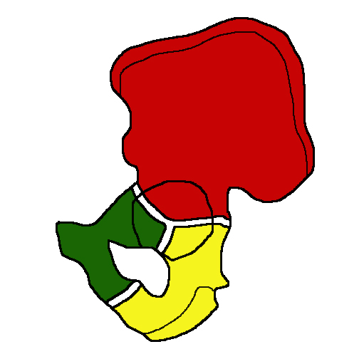 Innominate bone: Drawing demonstrating the unfused tri-radiate cartilage (white) and the three ossification centers of the ilium (red), ischium (yellow), and pubis (green).
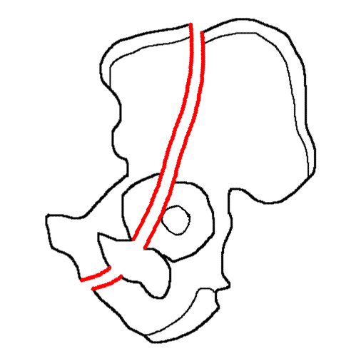
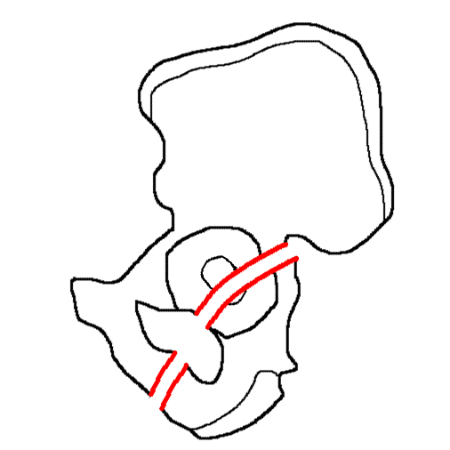
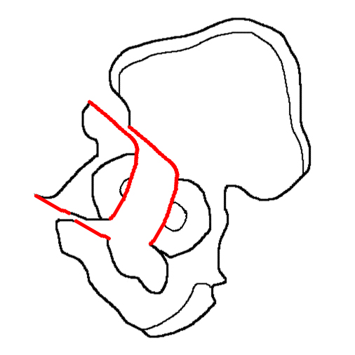
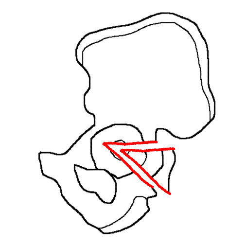
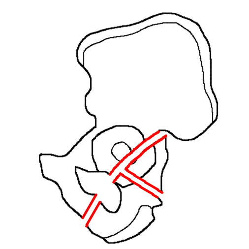
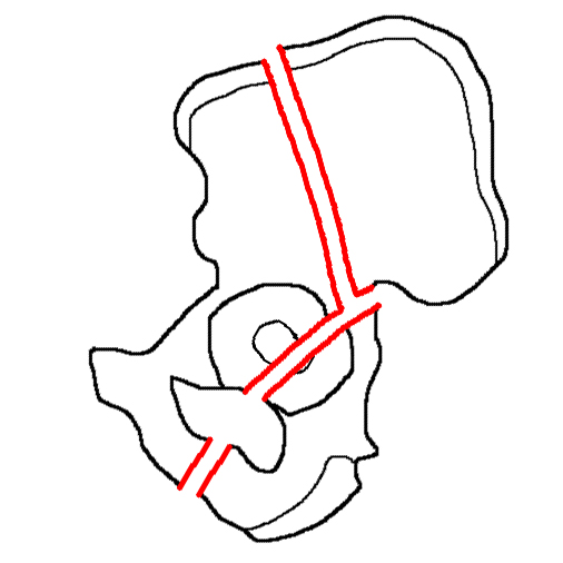
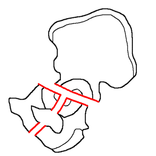
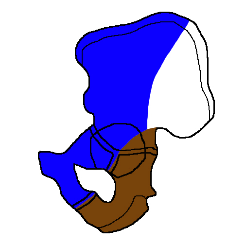 Innominate bone: Drawing demonstrating the anterior column (blue) and posterior column (brown) in an unfused innominate bone.

Lateral view of a 3D volume-rendered reconstruction of the innominate bone. Anterior column (blue) and posterior column (brown). The acetabulum is more than just the socket for the femoral head. It arises from, and is supported by, the innominate bone (os coxa), which forms from three ossification centers: the ilium, ischium, and pubis. These three segments join at the tri-radiate cartilage, a Y-shaped synchondrosis centered on the acetabulum. Complete fusion of the innominate bone occurs in the late teens. On lateral view, the innominate bone resembles the Greek letter lambda, with the longer limb forming the anterior column and the shorter, the posterior column of the acetabulum. These two columns serve as struts, mechanically representing the coalescence of bony trabeculae along lines of stress. Together, they transfer forces from the lower extremities through the hip and sacroiliac joints into the axial skeleton. Also important after injury, the columns function as anchors to which surgical hardware is attached. The anterior, or iliopubic, column is composed of the entire pubis and a large portion of the ilium, extending from the iliac crest down the iliac wing and through the superior obturator (pubic) ramus towards the pubic symphysis. Isolated injuries to the anterior column result from forces applied to the hip in external rotation. The posterior, or ilioischial, column is composed mainly of the ischium and a small part of the ilium. It extends from the posterior iliac body just below the angle of the greater sciatic notch down the ischial body into the inferior obturator (ischiopubic) ramus. Forces applied to the hip while in internal rotation result in posterior column injuries. Logically, the anterior and posterior acetabular walls are outward projections of their respective columns, joining superiorly at the tectum to form an articular cup for the femoral head. They add stability to the hip joint.
Ten acetabular fracture patterns in the Letournel-Judet classification system. Using this column principle, two orthopedists, Letournel and Judet, devised a comprehensive system for classifying acetabular fractures, consisting of ten injury patterns. Differentiating these patterns preoperatively is essential for choosing the appropriate surgical approach as no existing technique is able to expose both the anterior and posterior column intra-operatively. However, recognizing and remembering all ten patterns can be difficult, especially on cross-sectional imaging since the fracture patterns are named based on their appearance on the lateral view and not axial sections.
Thankfully, approximately 90% of all acetabular fractures fall into five patterns: isolated posterior wall, both column, transverse, transverse-posterior wall and t-type fractures. Moreover, the last two are actually variants of the transverse fracture, further narrowing the injuries down to three major patterns, which can easily be differentiated by the orientation of the fracture plane on axial CT images through the acetabular tectum.
|