of Acetabular Fractures:
Interactive CT Anatomy Learning Module | University of Washington
| Home | Quiz | Column Principle | Posterior Wall Fracture | Both Column Fracture | Transverse Fracture | Interactive Atlas | Summary | References |
University of Washington
Seattle, Washington |
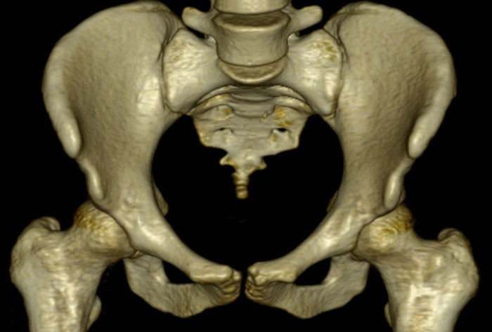
Welcome to the Interactive Learning Module of the Letournel-Judet Classification for Acetabular Fractures on CT |
|
The Letournel-Judet classification of acetabular fractures can be difficult to categorize on computed tomography (CT) because it was conceptualized from the lateral view of a hemipelvis. Proper treatment depends on accurate imaging assessment, as the injury pattern dictates the surgical approach. Our goals are to review the normal CT anatomy of the acetabulum and to help you recognize acetabular fractures according to the Letournel-Judet classification system. Mastery of this scheme will facilitate communication with orthopedists and enable appropriate and effective clinical management.
|