| Quick Start | Frontal | Maxillary | Ethmoid | Sphenoid | Interactive Atlas | Quiz |
University of Washington Department of Radiology Sung E. LoGerfo, M.D. |
Welcome to Interactive CT Sinus Anatomy |
|
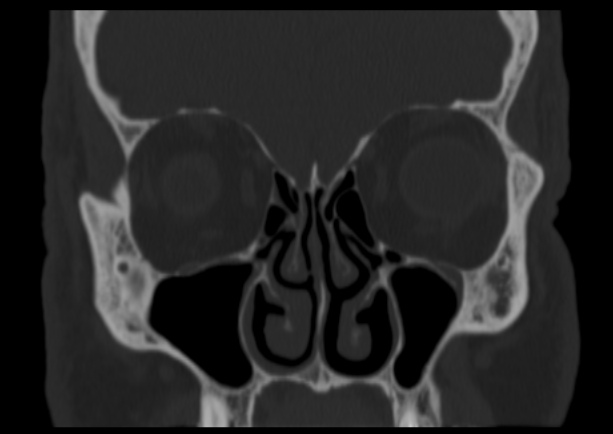
Imaging the paranasal sinuses is routine in clinical practice to evaluate for various sinus pathology, non-specific facial pain, and pre-operative planning for functional endoscopic sinus surgery (FESS), including post-operative follow-up. Our goal is to review the complex sinonasal anatomy, anatomic variants, mucociliary drainage pathways and inflammatory sinus disease. The teaching module highlights the frontal drainage sinus pathway, ostiomeatal unit and sphenoethmoidal recess with various cases illustrating involvement of the major drainage pathways in inflammatory sinus disease as well as clinical sequela of inflammatory sinus diesease. This module allows for interactive scrolling through normal sinuses in 3 planes, and includes labeled static images which highlight pertinent sinus anatomy. The user will be able to self-quiz at the end of the module to assess their knowledge of sinus anatomy, anatomic variants, mucociliary drainage patterns and inflammatory sinus disease. |