| Home | Frontal | Maxillary: Normal | Maxillary: Abnormal | Ethmoid | Sphenoid | Interactive Atlas | Quiz |
Maxillary Sinus: Normal Anatomy & Variants |
|
The maxillary sinuses usually develop symmetrically. The maxillary sinus ostium drains into the infundibulum which joins the hiatus semilunaris and drains into the middle meatus. The anterior ostiomeatal unit (OMU) is comprised of the frontal sinus ostium, frontal sinus drainage pathway (FSDP), maxillary sinus ostium, infundibulum, and middle meatus. These important structures connect the frontal, anterior ethmoid and maxillary sinuses.
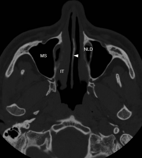
Axial image of the maxillary sinuses at the level of the nasal septum marked by arrowhead. (MS: maxillary sinus, NLD: nasolacrimal duct, IT: inferior turbinate)
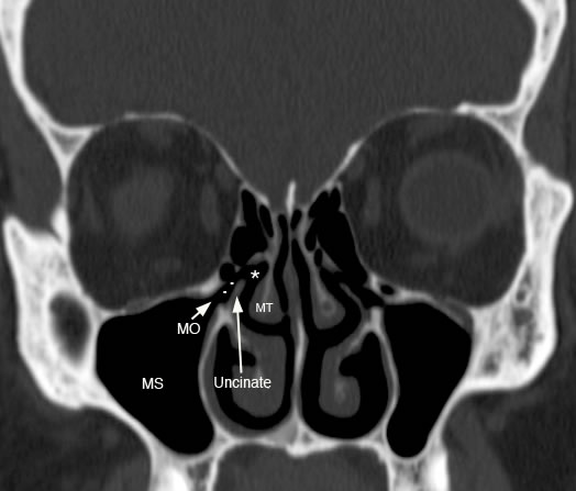
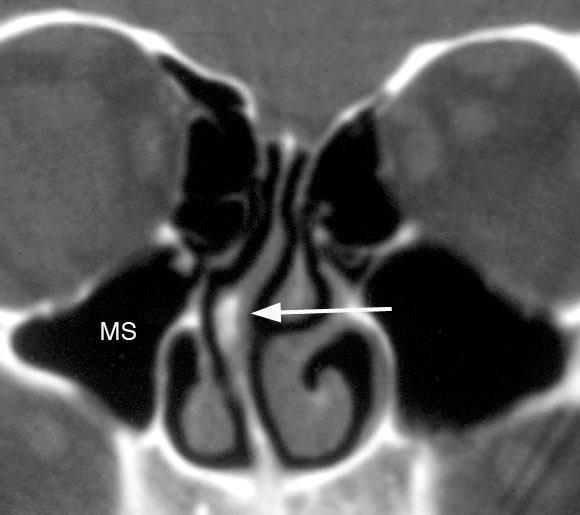
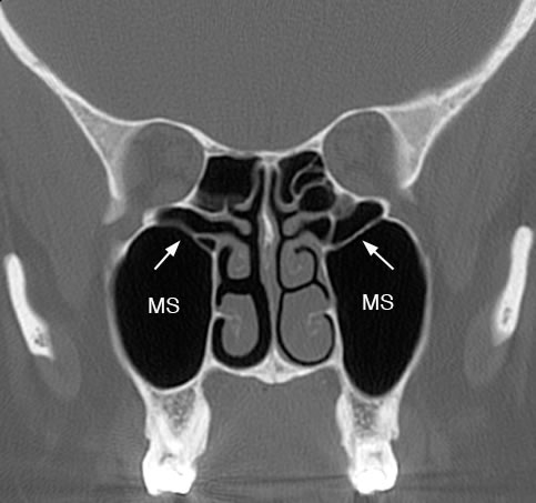
Coronal image with arrow pointing to maxillary sinus ostium (MO) with (..) illustrating the infundibulum joining the hiatus semilunaris (*). (MS: maxillary sinus, MT: middle turbinate)
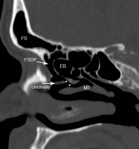
Sagittal image showing hiatus semilunaris (*) with uncinate process and ethmoid bulla (EB) superiorly. (MT: middle turbinate, FS: frontal sinus, FSDP: frontal sinus drainage pathyway)
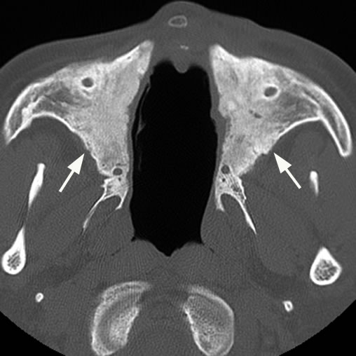
Axial images with arrows showing hypoplastic maxillary sinuses. The nasal septum is also absent. A septum can be absent due to either congenital or acquired (surgery, cocaine abuse or Wegener's granulomatosis) disorders.
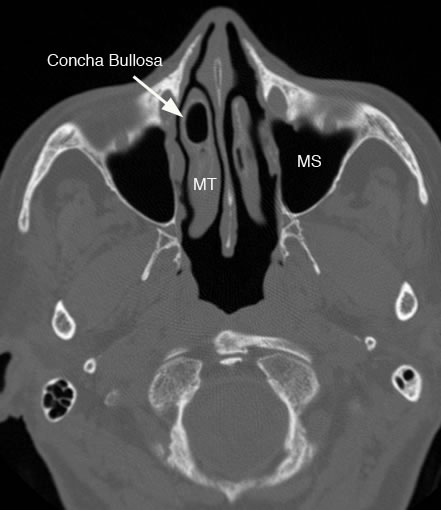
Axial image with arrow showing pneumatization of the middle turbinate (MT) also known as a concha bullosa which can potentially narrow the middle meatus. (MS: maxillary sinus)
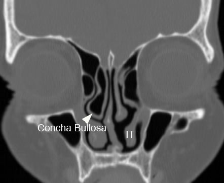
Coronal image with arrowhead showing concha bullosa of middle turbinate. Concha bullosa can potentially obstruct the maxillary sinus drainage pathway by narrowing the infundibulum and middle meatus.
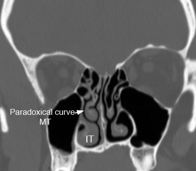
Coronal image with arrow pointing to paradoxical curvature of the middle turbinate (MT) with convexity of the bone directed laterally. Paradoxical curvature can potentially narrow or obstruct the infundibulum or middle meatus.
Coronal image with arrow pointing to nasal spur and septal deviation which if severe enough can narrow or compress the middle turbinate laterally and the middle meatus. (MS: maxillary sinus)
Coronal image with arrows illustrating thin bony septum in the maxillary sinuses (MS). |