| Home | Frontal: Normal | Frontal: Abnormal | Maxillary | Ethmoid | Sphenoid | Interactive Atlas | Quiz |
| Frontal Sinus: Normal Anatomy & Variants |
|
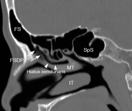
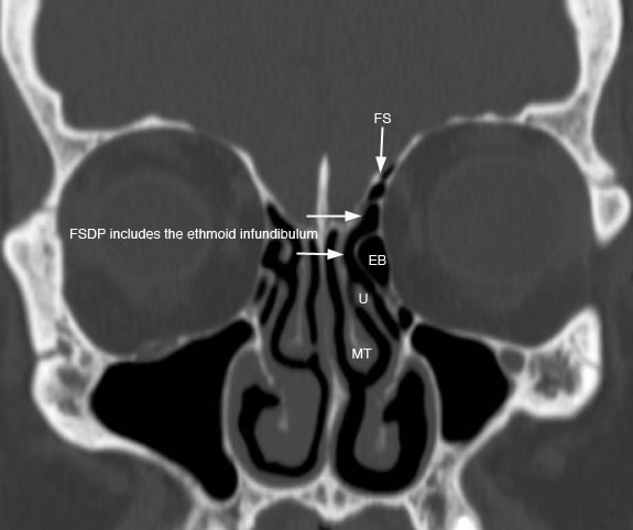
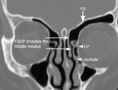
The frontal sinuses can have variable drainage depending on the anatomy of the frontal sinus drainage pathway (FSDP). The frontal sinuses have a superior and inferior compartment of the FSDP. The frontal sinus ostium drains into the superior compartment which then communicates directly with the inferior compartment. The inferior compartment is a narrow space that either is formed by the ethmoid infundibulum or middle meatus depending on the anterior attachment of the uncinate process. If the anterior uncinate process attaches superiorly to the skull base, then the inferior compartment of the FSDP is the ethmoid infundibulum which then communicates with the middle meatus via the hiatus semilunaris. If the anterior uncinate process attaches to the lamina papyracea, then the inferior compartment of the FSDP is the middle meatus.
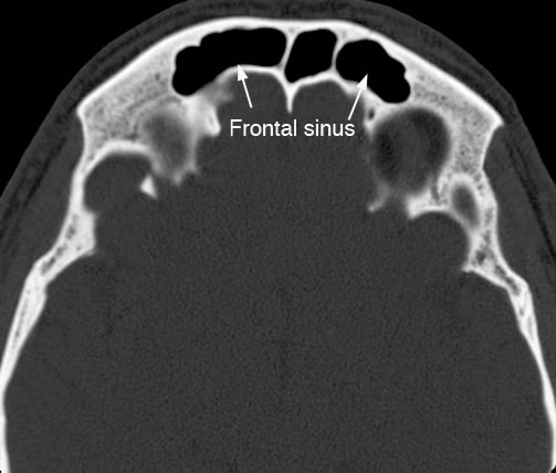
Axial image with arrows pointing to the frontal sinuses.
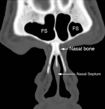
Coronal image of frontal sinuses (FS).
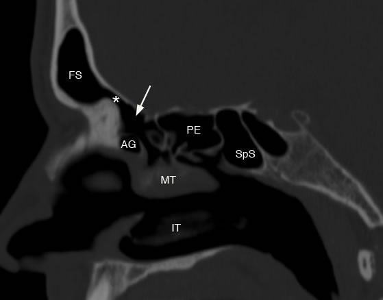
Sagittal image shows frontal sinus ostium (*) and arrow pointing to the superior compartment of the FSDP. (FS: frontal sinus, AG: agger nasi, PE: posterior ethmoid, SpS: sphenoid sinus, MT: middle turbinate, IT: inferior turbinate)
Sagittal image with arrows demonstrating frontal sinus drainage pathway and hiatus semilunaris which drains to middle meatus. (FS: frontal sinus, SpS: sphenoid sinus, MT: middle turbinate, IT: inferior turbinate)
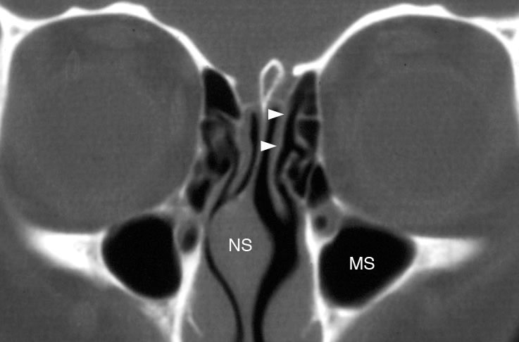
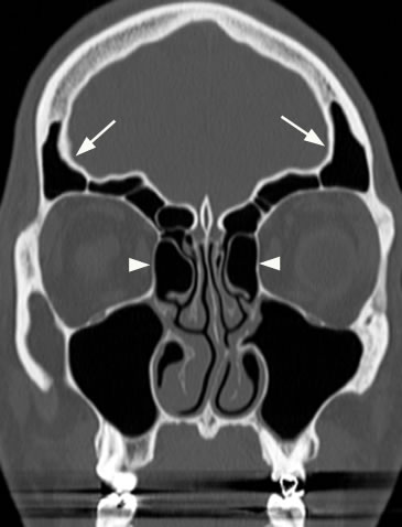
Coronal image with arrowheads demonstrating frontal sinus recess. (NS: nasal septum, MS: maxillary sinus)
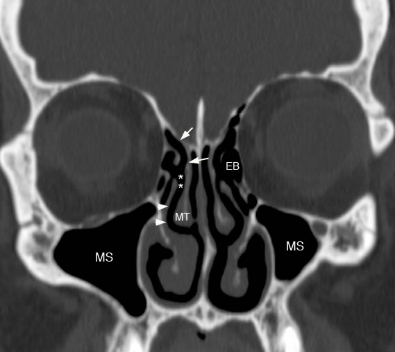
Coronal image demonstrating frontal recess (arrows), hiatus semilunaris (*) and middle meatus (arrowheads). (EB: ethmoid bulla, MT: middle turbinate, MS: maxillary sinus)
Coronal image with arrows demonstrating the inferior compartment of FSDP draining to ethmoid infundibulum because anterior process of uncinate attaches to skull base. (FS: frontal sinus, EB: ethmoid bulla, U: uncinate, MT: middle turbinate)
Coronal image with arrows demonstrating the inferior compartment of the FSDP includes the middle meatus because the anterior uncinate process attaches to the lamina papyracea. (FS: frontal sinus, LP: lamina papyracea)
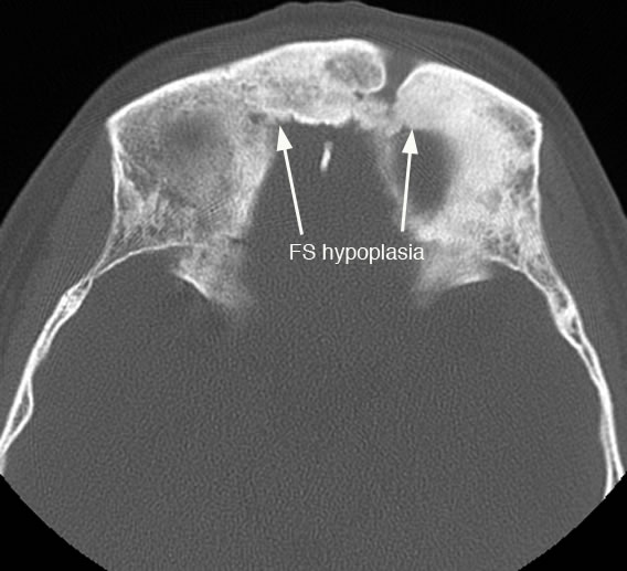
Axial image with arrows demonstrating hypoplastic frontal sinuses (FS).
Coronal image with arrows demonstrating overly pneumatized frontal sinuses. Also note enlarged ethmoid bulla air cells (arrowheads).
Coronal image with arrow pointing to pneumatized crista galli. Pneumatized crista galli may communicate with the frontal recess and can potentially obstruct the frontal sinus ostium. Incidentally noted is a tripod fracture involving the left maxillary sinus. |