| Home | Frontal: Normal | Frontal: Abnormal | Maxillary | Ethmoid | Sphenoid | Interactive Atlas | Quiz |
Frontal Sinus: Inflammatory Sinus Disease and Sequela |
|
Frontal sinus inflammatory disease can occur in isolation due to involvement of the ostium and frontal recess or as part of the anterior ostiomeatal complex (OMC) pattern which involves the frontal sinus drainage pathway, anterior ethmoid and maxillary sinuses. Inflammatory frontal sinus disease can result in mucus retention cysts, mucoceles or intracranial extension. Occasionally a benign osteoma can be present, and if large enough can obstruct the ostium or extend intracranially. Osteomas most commonly occur in the frontal sinuses.
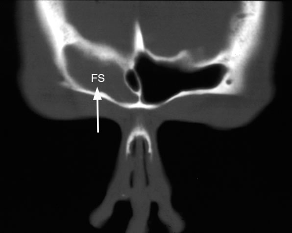
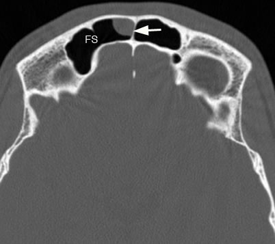
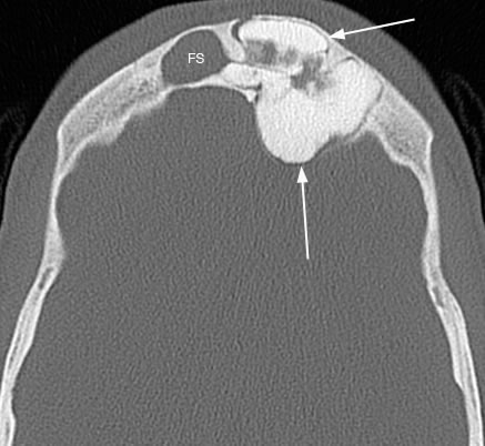
Coronal image with arrow pointing to isolated right frontal sinus (FS) disease.
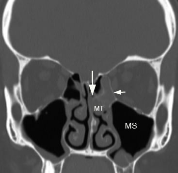
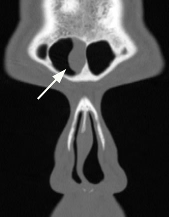
Coronal image illustrating sinus disease involving the anterior ethmoid sinus (arrows) and maxillary sinus (MS). Pattern of sinus disease indicates involvement of the anterior ostiomeatal unit. (MT: middle turbinate)
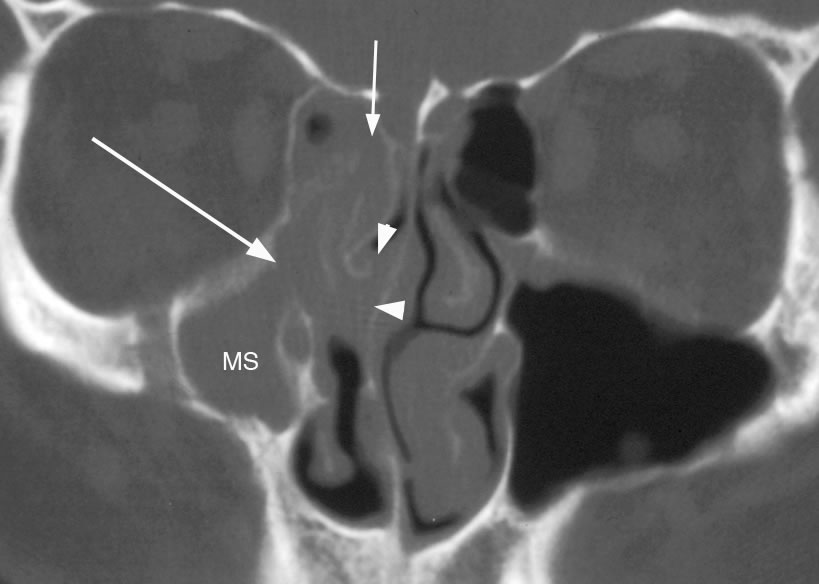
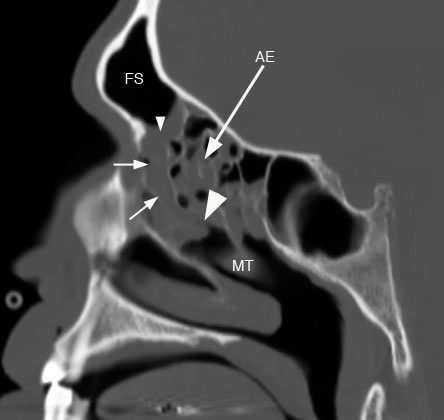
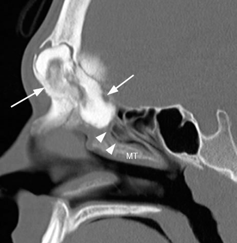
Coronal image demonstrating more diffuse anterior ostiomeatal sinus disease. Small arrow showing involvement of frontal recess and anterior ethmoid region, long arrow pointing to maxillary sinus ostium and infundibulum and arrowheads showing involvement of the hiatus semilunaris. Right maxillary sinus (MS) is hypoplastic. Sagittal image showing anterior ostiomeatal sinus disease involving the frontal sinus ostium (small arrowhead), frontal sinus drainage pathway (small arrows), hiatus semilunaris (large arrowhead) and anterior ethmoid sinus (AE). (FS: frontal sinus, MT: middle turbinate)
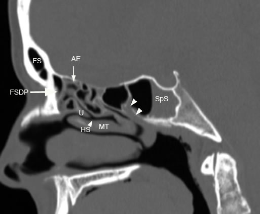
Sagitttal image demonstrating both anterior and posterior ostiomeatal sinus disease. There is mucosal thickening involving the FSDP, AE and sphenoid sinus ostium and sphenoethmoidal recess. (FS: frontal sinus, FSDP: frontal sinus drainage pathway, U: uncinate, HS: hiatus semilunaris, AE: anterior ethmoid, MT: middle turbinate, SpS: sphenoid sinus, double arrowheads: sphenoid sinus ostium and sphenoethmoidal recess)
Axial image of the frontal sinuses (FS) with arrow pointing to a mucus retention cyst along the non-dependant right frontal sinus air cell.
Coronal image of the frontal sinuses with large arrow pointing to mucus retention cyst.
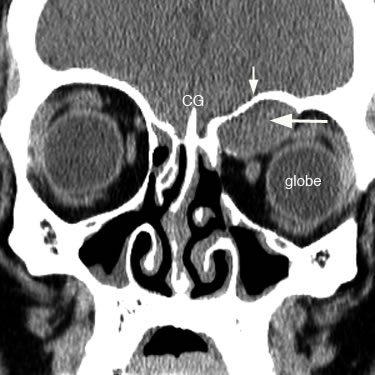
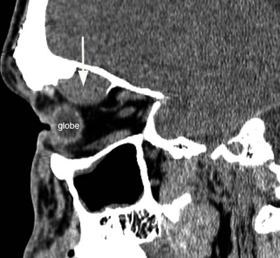
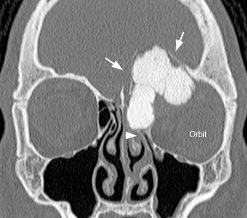
Coronl image with small arrow pointing to superior orbital roof and large arrow pointing to expansile frontal sinus mucocele extending into the orbit causing inferolateral displacement of the left globe. (CG: crista galli)
Sagittal image with arrow demonstrating large frontal sinus mucocele with orbital extension causing inferior displacement of the globe.
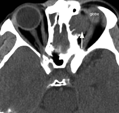
Axial image with arrow pointing to the expansile left frontal mucocele that has encroached into the orbit and is displacing the globe.
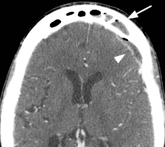
Axial post-contrast image shows arrowhead pointing to the peripherally enhancing intracranial extra-axial fluid collection containing small foci of gas which communicates with the left frontal sinus disease marked by the arrow. This patient developed an epidural abscess related to his frontal sinus disease.
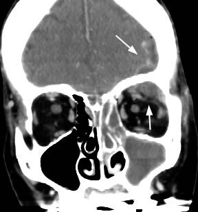
Coronal post-contrast image with arrows demonstrating large epidural abscess with both intracranial and orbital extension in a different patient. Also note the extensive unilateral sinus disease.
Axial image with arrows pointing to large left frontal sinus osteoma extending intracranially and obstructing the contralateral right frontal sinus (FS).
Coronal image with arrows pointing to the large frontal sinus osteoma extending intracranially and into the frontal recess. Note the inferior displacement of the left globe.
Sagittal image with arrows pointing to the large frontal osteoma extending intracranially and into the frontal recess. Arrowheads point to the hiatus semilunaris. (MT: middle turbinate) |