| Home | Frontal | Maxillary | Ethmoid | Sphenoid: Normal | Sphenoid: Abnormal | Interactive Atlas | Quiz |
Sphenoid Sinus: Normal Anatomy & Variants |
|
The sphenoid sinuses are highly variable in their configuration. Pneumatization can extend into the greater sphenoid wing resulting in lateral recesses. Additionally, pneumatization can also involve the posterior orbital wall, pterygoid processes, and lesser sphenoid wing. Important neighboring structures include the foramen rotundum, vidian canal, optic canal and internal carotid artery. The sphenoid sinus drains via the ostium into the sphenoethmoidal recess.
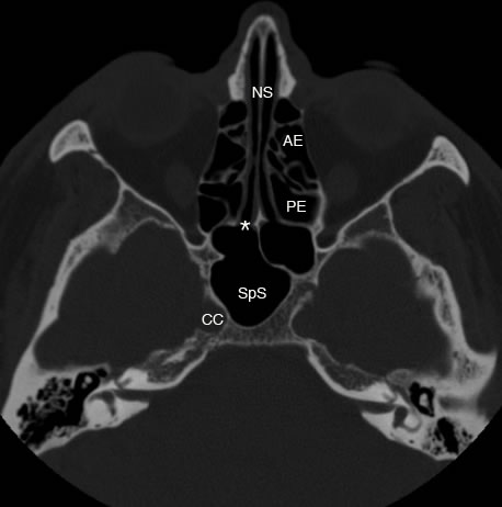
Axial image shows sphenoid sinus (SpS) and the sphenoethmoidal recess marked by the (*). (AE: anterior ethmoid, PE: posterior ethmoid, CC: carotid canal, NS: nasal septum)
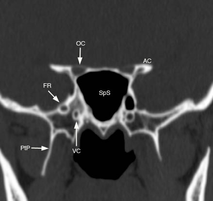
Coronal image of the sphenoid sinus (SpS) and neighboring structures. (FR: foramen rotundum, VC: vidian canal, OC: optic canal, AC: anterior clinoid, PtP: pterygoid plate)
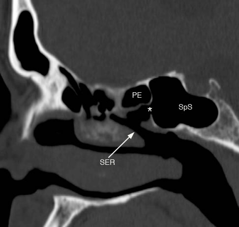
Sagittal image showing the sphenoid sinus (SpS) with sinus ostium (*) and arrow demonstrating the sphenoethmoidal recess (SER). (PE: posterior ethmois sinus)
Axial image with large arrows pointing to pneumatized lateral recesses of the sphenoid sinus (SpS). (FO: foramen ovale, FS: foramen spinosum)
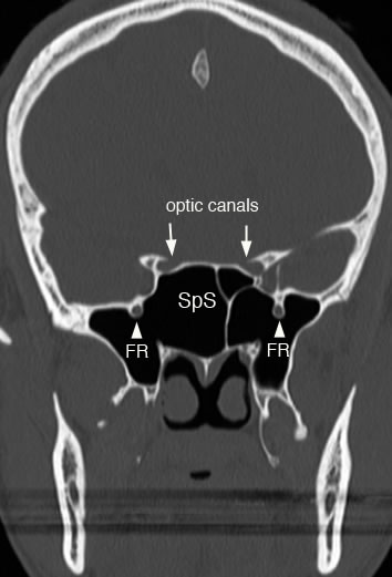
Coronal image showing pneumatized lateral recesses of sphenoid sinus (SpS) and foramen rotundum (FR) bulging into the sinus. Arrows point to optic canals superior to sphenoid sinus and medial to anterior clinoid processes.
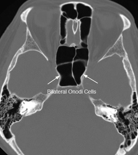
Axial image with arrows showing bilateral onodi air cells. Onodi air cells represent contigous extension of the posterior ethmoid air cells into the sphenoid sinus and are closely associated with the optic nerve. Due to their close association with the optic nerve the nerve can be at increased risk of injury during sinus surgery.
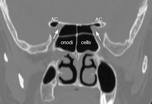
Coronal image showing bilateral onodi air cells with pneumatized anterior clinoid processes (AC) and arrows pointing to the optic canals.
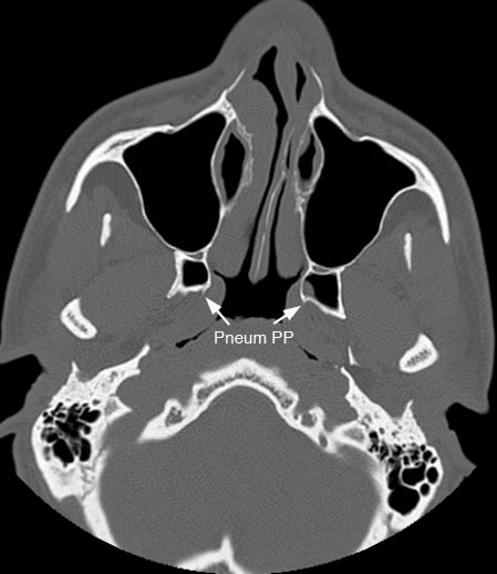
Axial image with arrows pointing to pneumatized pterygoid plates (PP).
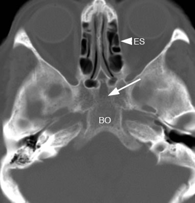
Axial image with arrow pointing to hypoplastic sphenoid sinus. (BO: basi-occiput, ES: ethmoid sinuses) |