| Home | Frontal | Maxillary | Ethmoid | Sphenoid: Normal | Sphenoid: Abnormal | Interactive Atlas | Quiz |
Sphenoid Sinus: Inflammatory Sinus Disease and Sequela |
|
Sphenoid sinuses drain via their ostium to the sphenoethmoidal recess and nasopharynx. Sphenoethmoidal recess inflammatory sinus disease pattern is usually due to either obstruction of the sinus in isolation or in conjunction with the posterior ethmoidal air cells. Mucoceles are not common in the sphenoid sinus but can occur and rarely sphenoid sinus disease can result in cavernous sinus thrombosis or intracranial extension.
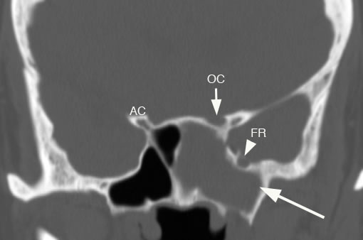
Coronal image of the sphenoid sinuses with arrow pointing to isolated left sphenoid sinus disease. Again notice the close anatomic proximity to the optic canal (OC) and foramen rotundum (FR). (AC: anterior clinoid process)
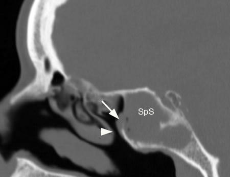
Sagittal image showing sphenoid sinus disease (SpS) with arrow showing obstructed sinus ostium and arrowhead pointing to sphenoethmoidal recess.
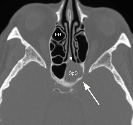
Axial image with large arrow showing left sphenoid sinus (SpS) disease with ostruction of the ostium (arrowhead). (EB: ethmoid bulla)
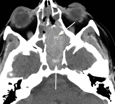
Axial image shows arrows pointing to a large expansile mass in the sphenoid sinus (SpS) extending into the posterior ethmoid sinus (PE) which was due to a large sphenoid sinus mucocele. (AE: anterior ethmoid sinus)
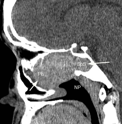
Sagittal image demonstrating large expansile sphenoid sinus (SpS) mucocele extending into the posterior ethmoid sinus (PE) and is being displaced anteriorly. (NP: nasopharynx)
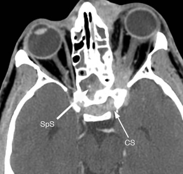
Axial post-contrast image demonstrating sphenoid sinus disease (SpS) with cavernous sinus thrombosis (CS) and orbital involvement. There is also ethmoid sinus disease.
Coronal post-contrast image showing sphenoid sinus disease (SpS) and cavernous sinus thrombosis (CS). |