| Scanning Notes for Biceps Tendon: The biceps tendon can be seen in transverse orientation by placing the back of the patient's hand on their ipsilateral thigh while seated, allowing 90 degree flexion of the elbow. The transducer is placed initially in axial position, allowing visualization of the greater and lesser tuberosities with the intervening biceps tendon and tendon sheath. Due to anisotropic effects, visualizing the tendon itself in this position can be difficult, as it tends to remain somewhat hypoechoic. This should not be mistaken for a tendon tear. Correlation with longitudinal images obtained with the transducer oriented sagitally along the tendon can alleviate this pitfall, as the tendon fibers should be clearly visible in this orientation. Fluid surrounding the biceps tendon within the tendon sheath is a fairly sensitive indicator of glenohumeral pathology, though as this space communicates with the shoulder capsule, such pathology is not necessarily limited to the biceps tendon and may represent a sign of synovitis or adhesive capsulitis. |
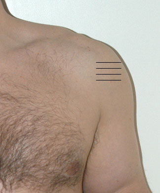
| Supraspinatus | Long | Trans |
| Infraspinatus | Long | Trans |
| Subscapularis | Long | Trans |
| Biceps Tendon | Long | Trans |