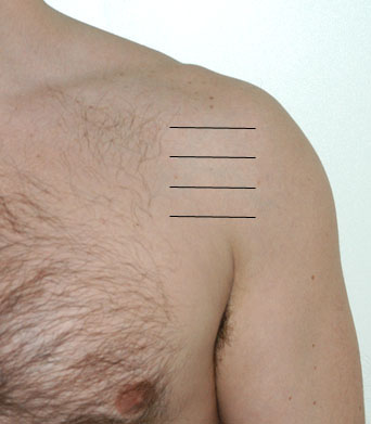
| Scanning Notes for Subscapularis: The subscapularis tendon is imaged in the same position as the biceps tendon, with the transducer placed more medially. Slight external rotation of the glenohumeral joint is helpful to elongate the tendon and allow more sensitive evaluation for tears. Images obtained during dynamic maneuvers in which the radiologist performs passive internal and external rotation of the shoulder with the elbow flexed 90 degrees helps to assess normal motion of the tendon, and allows further evaluation for tendon tears which will typically become more conspicuous with external rotation. Dislocation of the biceps tendon medially from the bicepital groove, indicative of full thickness tear of the transverse ligament of the supscapularis, may also be inapparent until passive external rotation. |

