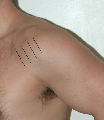
| Scanning Notes for Supraspinatus: These images are obtained in the modified Crass position, in which the patient extends and externally rotates the glenohumeral joint by placing the palm of his or her hand on the posterior aspect of the ipsilateral iliac wing and projects the flexed elbow joint posteriorly. This allows better visualization of the supraspinatus tendon, as in this position it will extend lateral to the acromion. Rotator cuff tears occur most commonly in this tendon, and are usually seen as hypoechoic defects in the contour of the tendon. Partial thickness tears can involve either the superficial (bursal) or deep (articular) surfaces of the tendon, and may be seen as either a mixed hypo- and hyperechoic focus or a solely hypoechoic focus extending to either tendon surface. Tears usually occur in the region of the critical zone, an area of relative avascularity within the tendon approximately 1 cm proximal to its insertion. |

